Explore
 Validate
 Learn
Learn Western blot
 Immunohistochemistry
Antibody data
- Antibody Data
- Antigen structure
- References [4]
- Comments [0]
- Validations
- Western blot [1]
- Immunocytochemistry [1]
- Other assay [1]
Submit
Validation data
Reference
Comment
Report error
- Product number
- MA1-10892 - Provider product page

- Provider
- Invitrogen Antibodies
- Product name
- HSP90 beta Monoclonal Antibody (H90-10)
- Antibody type
- Monoclonal
- Antigen
- Purifed from natural sources
- Description
- MA1-10892 detects 90 kDa Hsp90 beta in all reactive species except chicken where it detects both alpha and beta isoforms. MA1-10892 has been successfully used in Western blot and immunohistochemistry procedures. By Western blot, MA1-10892 is specific for the ~90 kDa Heat Shock Protein 90 protein. MA1-10892 immunogen is purified human Hsp90 beta.
- Reactivity
- Human, Mouse, Rabbit
- Host
- Mouse
- Isotype
- IgG
- Antibody clone number
- H90-10
- Vial size
- 100 µg
- Concentration
- 1.3 mg/mL
- Storage
- -20° C, Avoid Freeze/Thaw Cycles
Submitted references The Alpha Isoform of Heat Shock Protein 90 and the Co-chaperones p23 and Cdc37 Promote Opioid Anti-nociception in the Brain.
A C-terminal HSP90 inhibitor restores glucocorticoid sensitivity and relieves a mouse allograft model of Cushing disease.
GCUNC45 is the first Hsp90 co-chaperone to show alpha/beta isoform specificity.
The hsp90-related protein TRAP1 is a mitochondrial protein with distinct functional properties.
Lei W, Duron DI, Stine C, Mishra S, Blagg BSJ, Streicher JM
Frontiers in molecular neuroscience 2019;12:294
Frontiers in molecular neuroscience 2019;12:294
A C-terminal HSP90 inhibitor restores glucocorticoid sensitivity and relieves a mouse allograft model of Cushing disease.
Riebold M, Kozany C, Freiburger L, Sattler M, Buchfelder M, Hausch F, Stalla GK, Paez-Pereda M
Nature medicine 2015 Mar;21(3):276-80
Nature medicine 2015 Mar;21(3):276-80
GCUNC45 is the first Hsp90 co-chaperone to show alpha/beta isoform specificity.
Chadli A, Felts SJ, Toft DO
The Journal of biological chemistry 2008 Apr 11;283(15):9509-12
The Journal of biological chemistry 2008 Apr 11;283(15):9509-12
The hsp90-related protein TRAP1 is a mitochondrial protein with distinct functional properties.
Felts SJ, Owen BA, Nguyen P, Trepel J, Donner DB, Toft DO
The Journal of biological chemistry 2000 Feb 4;275(5):3305-12
The Journal of biological chemistry 2000 Feb 4;275(5):3305-12
No comments: Submit comment
Supportive validation
- Submitted by
- Invitrogen Antibodies (provider)
- Main image

- Experimental details
- Western blot analysis was performed on whole cell extracts (30 µg lysate) of A-431 (Lane 1), U-87 MG (Lane 2), HEK 293T (Lane 3), HeLa (Lane 4), K-562 (Lane 5) and NIH/3T3 (Lane 6). The blot was probed with Anti-HSP90 Monoclonal Antibody (H90-10) (Product # MA1-10892, 1µg/mL dilution) and detected by chemiluminescence using Goat anti-Mouse IgG (H+L) Superclonal™ Secondary Antibody, HRP conjugate (Product # A28177, 0.25 µg/mL, 1:4000 dilution). A 80 kDa band corresponding to HSP90 was observed across the cell lines tested.

Supportive validation
- Submitted by
- Invitrogen Antibodies (provider)
- Main image
- Experimental details
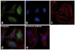
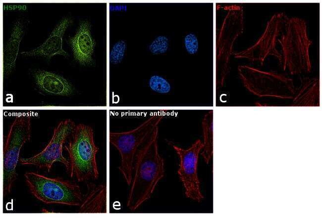
- Immunofluorescence analysis of HSP90 was performed using 70% confluent log phase HeLa cells. The cells were fixed with 4% paraformaldehyde for 10 minutes, permeabilized with 0.1% Triton™ X-100 for 10 minutes, and blocked with 1% BSA for 1 hour at room temperature. The cells were labeled with HSP90 Monoclonal Antibody(H90-10) (Product # MA1-10892) at 5µg/mL in 0.1% BSA and incubated overnight at 4 degree and then labeled with Goat anti-Mouse IgG (H+L) Superclonal™ Secondary Antibody, Alexa Fluor® 488 conjugate (Product # A28175) at a dilution of 1:2000 for 45 minutes at room temperature (Panel a: green). Nuclei (Panel b: blue) were stained with SlowFade® Gold Antifade Mountant with DAPI (Product # S36938). F-actin (Panel c: red) was stained with Rhodamine Phalloidin (Product # R415, 1:300). Panel d represents the merged image showing cytoplasmic and nuclear localization. Panel e represents control cells with no primary antibody to assess background. The images were captured at 60X magnification.
Supportive validation
- Submitted by
- Invitrogen Antibodies (provider)
- Main image

- Experimental details
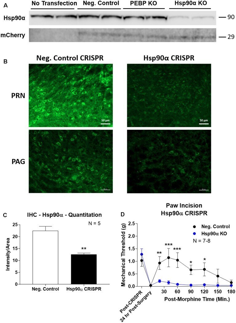
- Figure 3 CRISPR/Cas9 gene editing of Hsp90alpha in adult mouse brain. CRISPR constructs for all targets prepared and delivered as described in ""Materials and Methods"" section. All quantitative data reported as mean +- SEM. (A) Hsp90alpha CRISPR construct validated in 66.1 cells. Western blot shown with replicate wells of cells in each lane, with MW indicated for each target. Hsp90alpha protein levels reduced by ~90% only in the presence of Hsp90alpha-targeted CRISPR construct. Other constructs (Negative Control, PEBP) have no effect. mCherry protein levels are present in all transfected cells, demonstrating successful transfection of CRISPR DNA. (B) Hsp90alpha or negative control CRISPR delivered to CD-1 male mouse brains and analyzed for protein knockdown on day 10. Representative images shown from pontine reticular nucleus (PRN) and periaqueductal gray (PAG). Hsp90alpha (green signal) is present in cell bodies and dendritic trees, and the signal is reduced by CRISPR treatment. (C) Quantitation of all data from (B) performed as described in ""Materials and Methods"" section. Sample size of mice/group noted in graph. ** p < 0.01 vs. Negative Control group by Unpaired 2-Tailed t -test. Mice treated in one technical replicate, with the resulting tissue stained and analyzed in more than one technical replicate. CRISPR treatment reduced Hsp90alpha signal by 43.9%. (D) CRISPR-treated CD-1 male mice had paw incision surgery performed on day 9, with injection of 3.2 mg/kg morphine