Explore
 Validate
 Learn
Learn Western blot
 ELISA
ELISAAntibody data
- Antibody Data
- Antigen structure
- References [0]
- Comments [0]
- Validations
- Western blot [4]
- Immunohistochemistry [3]
- Flow cytometry [1]
Submit
Validation data
Reference
Comment
Report error
- Product number
- NB110-61640 - Provider product page

- Provider
- Novus Biologicals
- Proper citation
- Novus Cat#NB110-61640, RRID:AB_963937
- Product name
- Mouse Monoclonal HSP90 beta Antibody
- Antibody type
- Monoclonal
- Description
- Protein G purified. Detects 90 kDa protein. Detects HSP90 beta in all reactive species except in Chicken, where it detects both alpha and beta isoforms.
- Reactivity
- Human, Mouse, Rat, Canine, Chicken/Avian, Hamster, Rabbit
- Host
- Mouse
- Isotype
- IgG
- Vial size
- 0.2 mg
- Concentration
- 1 mg/ml
- Storage
- Store at 4C short term. Aliquot and store at -20C long term. Avoid freeze-thaw cycles.
No comments: Submit comment
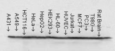
Supportive validation
- Submitted by
- Novus Biologicals (provider)
- Main image

- Experimental details
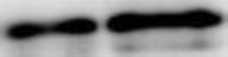
- Western Blot: HSP90AB1 Antibody (H90-10) [NB110-61640] - Human cell lysates from various cell lines showing detection of Hsp90 protein using Mouse Anti-Hsp90 Monoclonal Antibody, Clone H9010. 15 ug protein each lane. Block: 1.5% BSA for 30 minutes at RT. Primary antibody at 1:1000 for 2 hours at RT. Secondary antibody: Sheep Anti-Mouse IgG: HRP for 1 hour at RT.
- Submitted by
- Novus Biologicals (provider)
- Main image
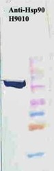
- Experimental details
- Western Blot: HSP90AB1 Antibody (H90-10) [NB110-61640] - Human HeLa cell lysates showing detection of Hsp90 protein using Mouse Anti-Hsp90 Monoclonal Antibody, Clone H9010 . Primary Antibody: Mouse Anti-Hsp90 Monoclonal Antibody at 1:1000. Secondary Antibody: HRP Goat Anti-Mouse.
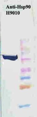
- Submitted by
- Novus Biologicals (provider)
- Main image

- Experimental details
- Western Blot: HSP90AB1 Antibody (H90-10) [NB110-61640] - Human Lysates showing detection of Hsp90 protein using Mouse Anti-Hsp90 Monoclonal Antibody, Clone H9010 . Primary Antibody: Mouse Anti-Hsp90 Monoclonal Antibody at 1:1000. Comparison of clone H9010 behavior with Hsp90 human beta (1) and Hsp90 human alpha (2). Courtesy of: David Toft, Mayo Clinic.

- Submitted by
- Novus Biologicals (provider)
- Main image
- Experimental details
- Western Blot: HSP90AB1 Antibody (H90-10) [NB110-61640] - HCC827 human non-small cell lung cancer cell line and A549 cells lysates. Image from verified customer review.

Supportive validation
- Submitted by
- Novus Biologicals (provider)
- Main image
- Experimental details
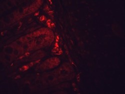
- Immunohistochemistry: HSP90AB1 Antibody (H90-10) [NB110-61640] - Mouse inflammed colon tissue. Fixation: Formalin. Primary antibody at 1:10000 for 12 hours at 4C. Secondary antibody: Alexa Fluor 555 Goat Anti-Mouse (red) at 1:5000 for 1 hour at RT. Localization: Inflammatory and epithelial mucosa. Magnification: 40x.
- Submitted by
- Novus Biologicals (provider)
- Main image
- Experimental details
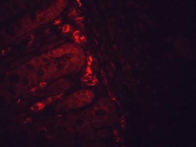
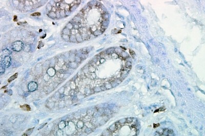
- Immunohistochemistry-Paraffin: HSP90AB1 Antibody (H90-10) [NB110-61640] - Cancerous human colon tissue.

- Submitted by
- Novus Biologicals (provider)
- Main image

- Experimental details
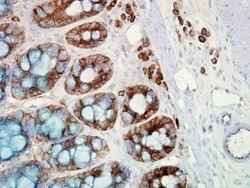
- Immunohistochemistry-Paraffin: HSP90AB1 Antibody (H90-10) [NB110-61640] - Tissue: inflamed colon. Species: Mouse. Fixation: Formalin. Primary Antibody: Mouse Anti-Hsp90 Monoclonal Antibody at 1:10000 for 12 hours at 4C. Secondary Antibody: Biotin Goat Anti-Mouse at 1:2000 for 1 hour at RT. Counterstain: Mayer Hematoxylin (purple/blue) nuclear stain at 200 ul for 2 minutes at RT. Localization: Inflammatory cells. Magnification: 40x.
Supportive validation
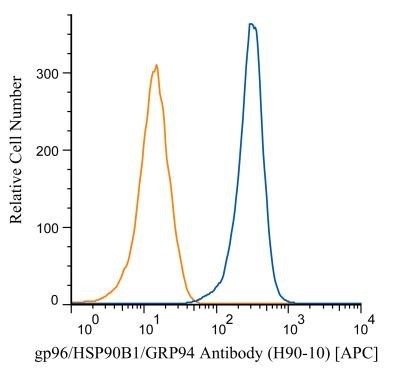
- Submitted by
- Novus Biologicals (provider)
- Main image
- Experimental details
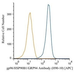
- Flow Cytometry: HSP90AB1 Antibody (H90-10) [NB110-61640] - An intracellular stain was performed on Jurkat cells with antibody NB110-61640APC (blue) and a matched isotype control (orange). Cells were fixed with 4% PFA and then permeablized with 0.1% saponin. An antibody dilution of 1:100 was used and cells were incubated for 30 minutes at room temperature. Image using the Allophycocyanin form of this antibody.