Explore
 Validate
 Learn
Learn Western blot
 ELISA
ELISA Immunocytochemistry
Antibody data
- Antibody Data
- Antigen structure
- References [0]
- Comments [0]
- Validations
- Immunocytochemistry [3]
- Immunohistochemistry [1]
Submit
Validation data
Reference
Comment
Report error
- Product number
- MA5-45155 - Provider product page

- Provider
- Invitrogen Antibodies
- Product name
- HSP90 alpha/beta Monoclonal Antibody (Hyb-K41220A), FITC
- Antibody type
- Monoclonal
- Antigen
- Recombinant full-length protein
- Description
- 1 µg/mL was sufficient for detection of HSP90 alpha beta in 20 µg of heat shocked HeLa cell lysate by colorimetric immunoblot analysis using Goat Anti-Mouse IgG:HRP as the secondary.|Detects 90kDa. Will detect both alpha (inducible) and beta (constitutively-expressed) forms.
- Reactivity
- Human, Mouse, Rat, Yeast
- Host
- Mouse
- Conjugate
- Green dye
- Isotype
- IgG
- Antibody clone number
- Hyb-K41220A
- Vial size
- 100 μg
- Concentration
- 1 mg/mL
- Storage
- 4°C, store in dark
No comments: Submit comment
Supportive validation
- Submitted by
- Invitrogen Antibodies (provider)
- Main image
- Experimental details
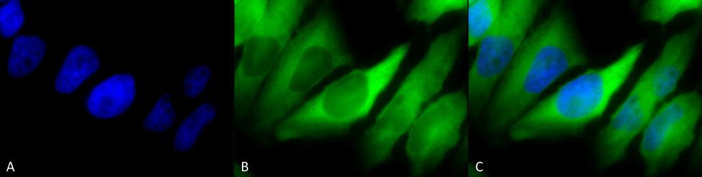
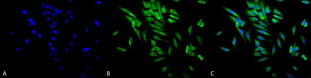
- Immunocytochemistry/Immunofluorescence analysis using human cervical cancer cells. Fixation involved 2% Formaldehyde for 20 min at RT. Samples were incubated with Hsp90 alpha/beta monoclonal antibody (Product # MA5-45155) at 1:100 for 12 hours at 4°C, followed by FITC Goat Anti-Mouse (green) at 1:200 for 2 hours at RT. Counterstain used was DAPI (blue) nuclear stain at 1:40,000 for 2 hours at RT. Localization: Cytoplasm. Melanosome. Magnification: 100x. (A) DAPI (blue) nuclear stain. (B) Anti-Hsp90 alpha/beta antibody. (C) Composite.
- Submitted by
- Invitrogen Antibodies (provider)
- Main image
- Experimental details
- Immunocytochemistry/Immunofluorescence analysis using human cervical cancer cells. Fixation involved 2% Formaldehyde for 20 min at RT. Samples were incubated with Hsp90 alpha/beta monoclonal antibody (Product # MA5-45155) at 1:100 for 12 hours at 4°C, followed by FITC Goat Anti-Mouse (green) at 1:200 for 2 hours at RT. Counterstain used was DAPI (blue) nuclear stain at 1:40,000 for 2 hours at RT. Localization: Cytoplasm. Melanosome. Magnification: 20x. (A) DAPI (blue) nuclear stain. (B) Anti-Hsp90 alpha/beta antibody. (C) Composite.
- Conjugate
- Green dye
- Submitted by
- Invitrogen Antibodies (provider)
- Main image
- Experimental details
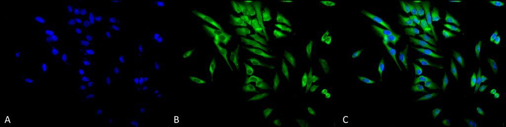
- Immunocytochemistry/Immunofluorescence analysis using human cervical cancer cells. Fixation involved 2% Formaldehyde for 20 min at RT. Samples were incubated with Hsp90 alpha/beta monoclonal antibody (Product # MA5-45155) at 1:100 for 12 hours at 4°C, followed by FITC Goat Anti-Mouse (green) at 1:200 for 2 hours at RT. Counterstain used was DAPI (blue) nuclear stain at 1:40,000 for 2 hours at RT. Localization: Cytoplasm. Melanosome. Magnification: 20x. (A) DAPI (blue) nuclear stain. (B) Anti-Hsp90 alpha/beta antibody. (C) Composite.
Supportive validation
- Submitted by
- Invitrogen Antibodies (provider)
- Main image
- Experimental details
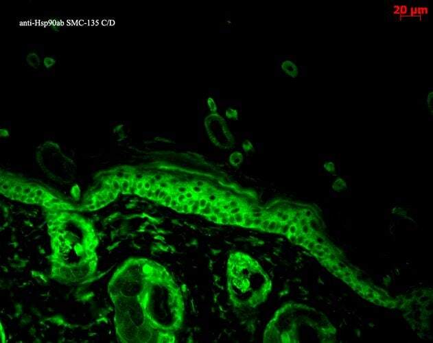
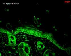
- Immunohistochemistry analysis using backskin. Fixation involved Bouins Fixative and paraffin-embedded. Samples were incubated with Hsp90 alpha monoclonal antibody (Product # MA5-45155) at 1:100 for 1 hour at RT, followed by FITC Goat Anti-Mouse (green) at 1:50 for 1 hour at RT. Localization: Epidermis.