Explore
 Validate
 Learn
LearnPA5-80339
antibody from Invitrogen Antibodies
Targeting: ANP32A
C15orf1, I1PP2A, LANP, MAPM, mapmodulin, PHAPI, PP32
 Western blot
 ELISA
ELISA Immunocytochemistry
Antibody data
- Antibody Data
- Antigen structure
- References [1]
- Comments [0]
- Validations
- Immunocytochemistry [3]
- Immunoprecipitation [1]
- Immunohistochemistry [2]
- Other assay [2]
Submit
Validation data
Reference
Comment
Report error
- Product number
- PA5-80339 - Provider product page

- Provider
- Invitrogen Antibodies
- Product name
- PHAP I Polyclonal Antibody
- Antibody type
- Polyclonal
- Antigen
- Recombinant full-length protein
- Description
- This product is preservative free. It is recommended to add sodium azide to avoid contamination (final concentration 0.05%-0.1%). This antibody has specificity for Human ANP32A/PHAP1.
- Reactivity
- Human
- Host
- Rabbit
- Isotype
- IgG
- Vial size
- 100 μL
- Concentration
- 1 mg/mL
- Storage
- Store at 4°C short term. For long term storage, store at -20°C, avoiding freeze/thaw cycles.
Submitted references Comparative analyses of alphaviral RNA:Protein complexes reveals conserved host-pathogen interactions.
Gebhart NN, Hardy RW, Sokoloski KJ
PloS one 2020;15(8):e0238254
PloS one 2020;15(8):e0238254
No comments: Submit comment
Supportive validation
- Submitted by
- Invitrogen Antibodies (provider)
- Main image

- Experimental details
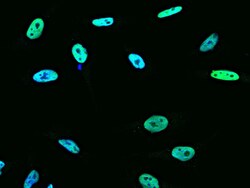
- Immunofluorescence staining of PHAP I in HeLa cells. Cells were fixed with 4% PFA, permeabilzed with 0.3% Triton X-100 in PBS, blocked with 10% serum, and incubated with PHAP I Polyclonal Antibody (Product # PA5-80339, 1:1,000) at 4°C overnight. Then cells were stained with the Alexa Fluor®488-conjugated Goat Anti-rabbit IgG secondary antibody (green) and counterstained with DAPI (blue). Positive staining was localized to nucleus.

- Submitted by
- Invitrogen Antibodies (provider)
- Main image
- Experimental details
- Immunofluorescence staining of PHAP I in HeLa cells. Cells were fixed with 4% PFA, permeabilzed with 0.3% Triton X-100 in PBS, blocked with 10% serum, and incubated with PHAP I Polyclonal Antibody (Product # PA5-80339, 1:1,000) at 4°C overnight. Then cells were stained with the Alexa Fluor®488-conjugated Goat Anti-rabbit IgG secondary antibody (green) and counterstained with DAPI (blue). Positive staining was localized to nucleus.

- Submitted by
- Invitrogen Antibodies (provider)
- Main image

- Experimental details
- Immunofluorescence staining of PHAP I in HeLa cells. Cells were fixed with 4% PFA, permeabilzed with 0.3% Triton X-100 in PBS, blocked with 10% serum, and incubated with PHAP I Polyclonal Antibody (Product # PA5-80339, 1:1,000) at 4°C overnight. Then cells were stained with the Alexa Fluor®488-conjugated Goat Anti-rabbit IgG secondary antibody (green) and counterstained with DAPI (blue). Positive staining was localized to nucleus.
Supportive validation
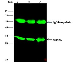
- Submitted by
- Invitrogen Antibodies (provider)
- Main image

- Experimental details
- PHAP I Immunoprecipitation using: Lane A: 0.5 mg Jurkat Whole Cell Lysate, Lane B: 0.5 mg Hela Whole Cell Lysate, Lane C: 0.5 mg HepG2 Whole Cell Lysate 4 µL with PHAP I Polyclonal Antibody (Product # PA5-80339) and 15 µL of 50 % Protein G agarose. Primary antibody: PHAP I Polyclonal Antibody, at 1:100 dilution. Secondary antibody: Dylight 800-labeled antibody to rabbit IgG (H+L), at 1:5,000 dilution. Developed using the Odyssey technique. Performed under reducing conditions. Predicted band size: 29 kDa. Observed band size: 29 kDa.

Supportive validation
- Submitted by
- Invitrogen Antibodies (provider)
- Main image

- Experimental details
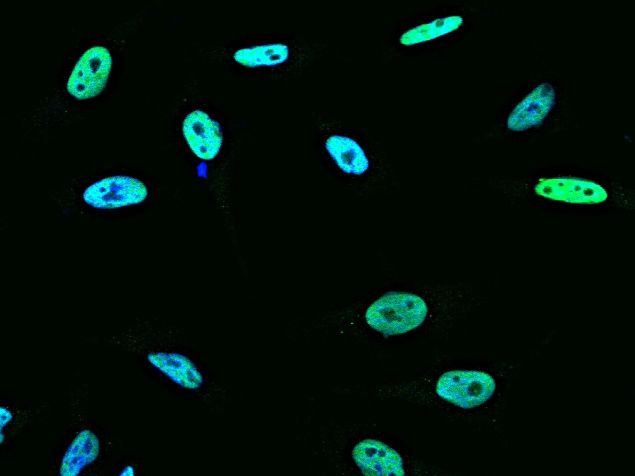
- Immunohistochemical staining of human PHAP I in human kidney with PHAP I Polyclonal Antibody (Product # PA5-80339, 1:1,000, formalin-fixed paraffin embedded sections).
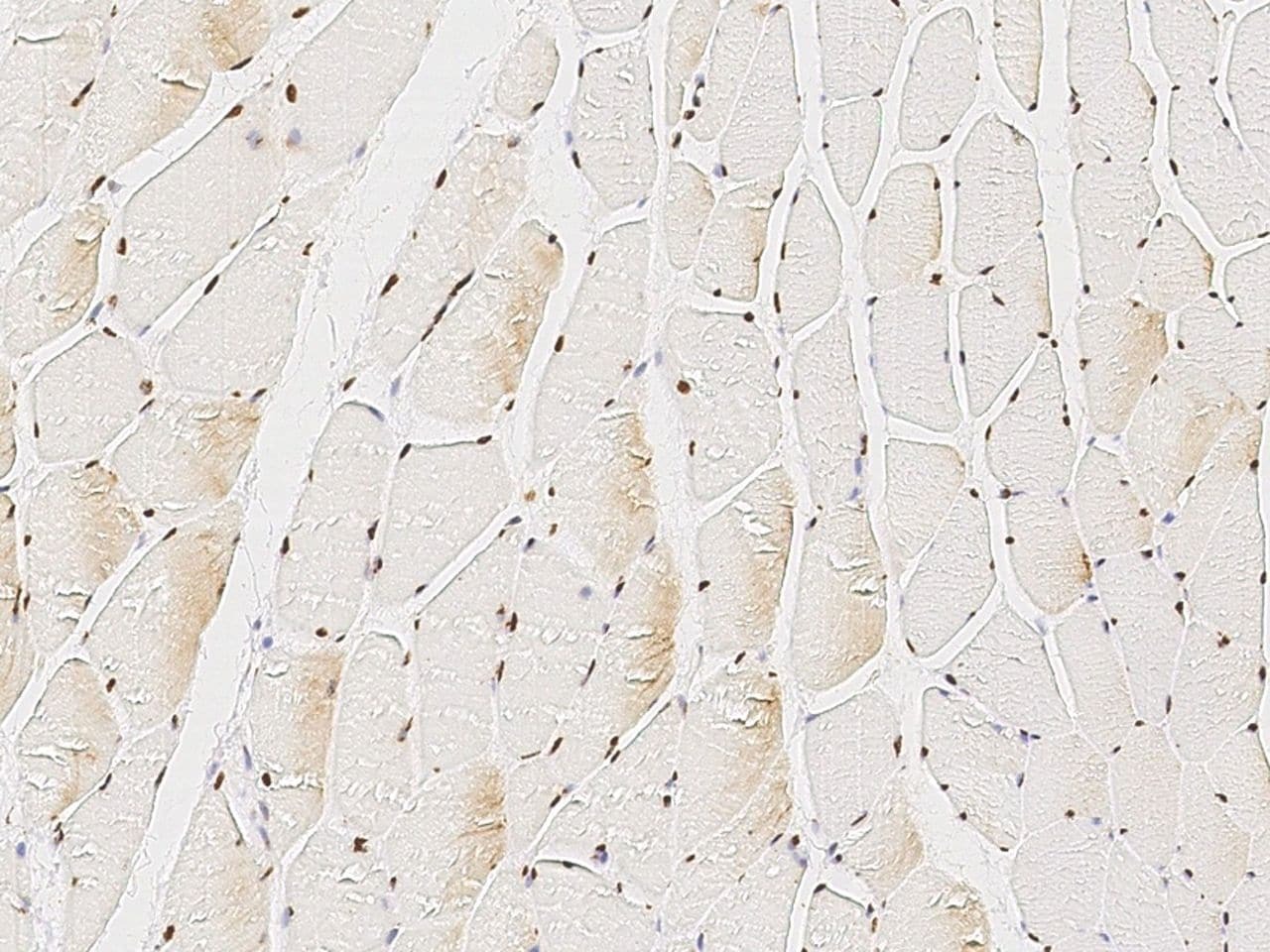
- Submitted by
- Invitrogen Antibodies (provider)
- Main image
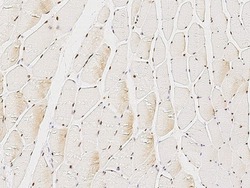
- Experimental details
- Immunohistochemical staining of human PHAP I in human skeletal muscle with PHAP I Polyclonal Antibody (Product # PA5-80339, 1:1,000, formalin-fixed paraffin embedded sections).
Supportive validation
- Submitted by
- Invitrogen Antibodies (provider)
- Main image
- Experimental details
- PHAP I Immunoprecipitation using: Lane A: 0.5 mg Jurkat Whole Cell Lysate, Lane B: 0.5 mg Hela Whole Cell Lysate, Lane C: 0.5 mg HepG2 Whole Cell Lysate 4 µL with PHAP I Polyclonal Antibody (Product # PA5-80339) and 15 µL of 50 % Protein G agarose. Primary antibody: PHAP I Polyclonal Antibody, at 1:100 dilution. Secondary antibody: Dylight 800-labeled antibody to rabbit IgG (H+L), at 1:5,000 dilution. Developed using the Odyssey technique. Performed under reducing conditions. Predicted band size: 29 kDa. Observed band size: 29 kDa.

- Submitted by
- Invitrogen Antibodies (provider)
- Main image
- Experimental details
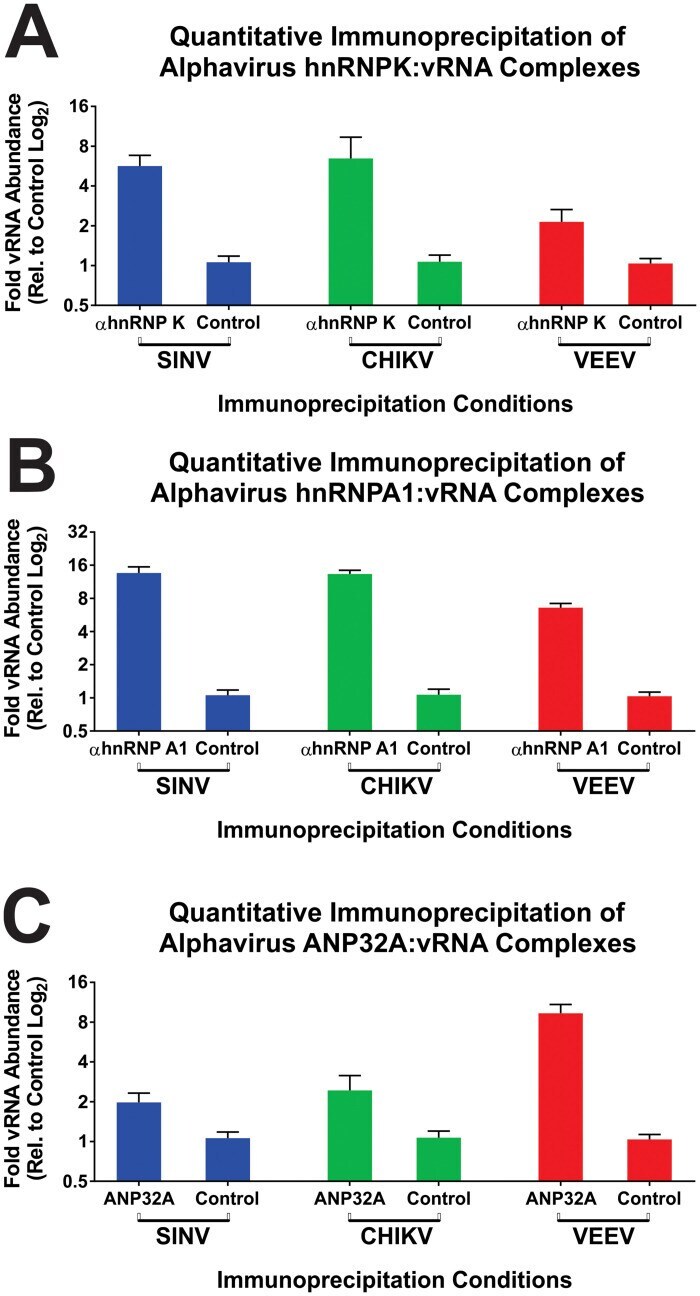
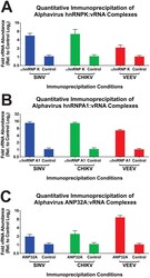
- Fig 9 The validation of select CLAMP identified common interactants. 293HEK cells were infected with SINV, CHIKV, or VEEV at an MOI of 10 PFU/Cell and processed as described in the materials and methods. Briefly, the infected cells were cross-linked prior to the generation of whole cell lysates. After clarification via centrifugation the cross-linked lysates were immunoprecipitated with either anti-hnRNP K (Panel A), anti-hnRNP A1 (Panel B), or anti-ANP32a antibodies (Panel C). Nonspecific control immunoprecipitations utilizing an anti-IL1 antibody were conducted in parallel. After immunoprecipitation the cross-links were reversed, and the co-immunoprecipitated RNAs were used as input materials for the synthesis of random hexamer primed cDNAs. The positive sense viral RNA species of SINV, CHIKV, and VEEV were then assayed using qRT-PCR and plotted as the fold abundance of the viral RNA relative to the control immunoprecipitations. The quantitative data shown is the means of three independent biological replicates, and the error bar represents the standard deviation of the means.