 Explore
 Validate
 Learn
Learn Western blot
 Immunocytochemistry
Antibody data
- Antibody Data
- Antigen structure
- References [3]
- Comments [0]
- Validations
- Western blot [5]
- Immunocytochemistry [1]
- Immunoprecipitation [1]
Submit
Validation data
Reference
Comment
Report error
- Product number
- GTX113834 - Provider product page

- Provider
- GeneTex
- Proper citation
- GeneTex Cat#GTX113834, RRID:AB_2037974
- Product name
- SET antibody
- Antibody type
- Polyclonal
- Reactivity
- Human, Mouse, Rat
- Host
- Rabbit
Submitted references Neuronal ceroid lipofuscinosis related ER membrane protein CLN8 regulates PP2A activity and ceramide levels.
SET protein overexpression contributes to paclitaxel resistance in MCF-7/S cells through PI3K/Akt pathway.
Proteomic analysis of the proteins that are associated with the resistance to paclitaxel in human breast cancer cells.
Adhikari B, De Silva B, Molina JA, Allen A, Peck SH, Lee SY
Biochimica et biophysica acta. Molecular basis of disease 2019 Feb 1;1865(2):322-328
Biochimica et biophysica acta. Molecular basis of disease 2019 Feb 1;1865(2):322-328
SET protein overexpression contributes to paclitaxel resistance in MCF-7/S cells through PI3K/Akt pathway.
Zhang W, Zheng X, Meng T, You H, Dong Y, Xing J, Chen S
Journal of drug targeting 2017 Mar;25(3):255-263
Journal of drug targeting 2017 Mar;25(3):255-263
Proteomic analysis of the proteins that are associated with the resistance to paclitaxel in human breast cancer cells.
Chen S, Dong Q, Hu S, Cai J, Zhang W, Sun J, Wang T, Xie J, He H, Xing J, Lu J, Dong Y
Molecular bioSystems 2014 Feb;10(2):294-303
Molecular bioSystems 2014 Feb;10(2):294-303
No comments: Submit comment
Enhanced validation
Supportive validation
- Submitted by
- GeneTex (provider)
- Enhanced method
- Genetic validation
- Main image
- Experimental details
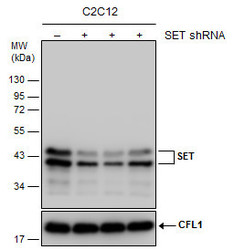
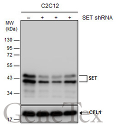
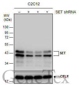
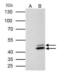
- Non-transfected (¡V) and transfected (+) C2C12 whole cell extracts (30 ?g) were separated by 12% SDS-PAGE, and the membrane was blotted with SET antibody (GTX113834) diluted at 1:1000.
Supportive validation
- Submitted by
- GeneTex (provider)
- Main image
- Experimental details
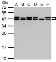
- SET antibody detects SET protein by Western blot analysis.A. 30 ?g A431 whole cell lysate/extract whole cell lysate/extract B. 30 ?g H1299 whole cell lysate/extract C. 30 ?g HeLa whole cell lysate/extract D. 30 ?g HepG2 whole cell lysate/extract E. 30 ?g Molt-4 whole cell lysate/extract F. 30 ?g Raji whole cell lysate/extract12 % SDS-PAGESET antibody (GTX113834) dilution: 1:1000 SET antibody detects SET protein by Western blot analysis.

- Submitted by
- GeneTex (provider)
- Main image
- Experimental details
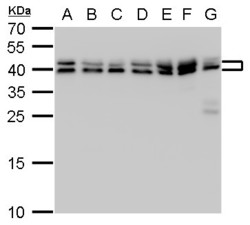
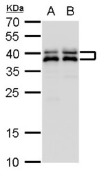
- SET antibody detects SET protein by western blot analysis.A. 30 £gg Neuro2A whole cell lysate/extract B. 30 £gg GL261 whole cell lysate/extract C. 30 £gg C8D30 whole cell lysate/extract D. 30 £gg NIH-3T3 whole cell lysate/extract E. 30 £gg BCL-1 whole cell lysate/extract F. 30 £gg Raw 264.7 whole cell lysate/extract G. 30 £gg C2Cl2 whole cell lysate/extract12 % SDS-PAGESET antibody (GTX113834) dilution: 1:1000

- Submitted by
- GeneTex (provider)
- Main image
- Experimental details
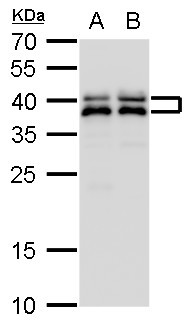
- SET antibody detects SET protein by western blot analysis.A. 30 £gg PC-12 whole cell lysate/extract B. 30 £gg Rat2 whole cell lysate/extract12 % SDS-PAGESET antibody (GTX113834) dilution: 1:1000
- Submitted by
- GeneTex (provider)
- Main image
- Experimental details
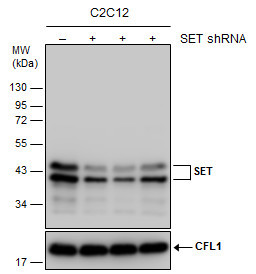
- Non-transfected (¡V) and transfected (+) C2C12 whole cell extracts (30 ?g) were separated by 12% SDS-PAGE, and the membrane was blotted with SET antibody (GTX113834) diluted at 1:1000.
Supportive validation
- Submitted by
- GeneTex (provider)
- Main image

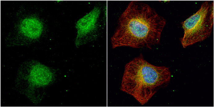
- Experimental details
- SET antibody detects SET protein at cytoplasm and nucleus by immunofluorescent analysis.Sample: HeLa cells were fixed in 4% paraformaldehyde at RT for 15 min.Green: SET protein stained by SET antibody (GTX113834) diluted at 1:500.Red: alpha Tubulin, a cytoskeleton marker, stained by alpha Tubulin antibody [GT114] (GTX628802) diluted at 1:1000.Blue: Hoechst 33342 staining.
Supportive validation
- Submitted by
- GeneTex (provider)
- Main image
- Experimental details
- SET antibody immunoprecipitates SET protein in IP experiments.IP samples: HeLa whole cell extractA. Control with 4 £gg of preimmune Rabbit IgGB. Immunoprecipitation of SET protein by 4 £gg SET antibody (GTX113834)10 % SDS-PAGEThe immunoprecipitated SET protein was detected by SET antibody (GTX113834) diluted at 1:500.[EasyBlot anti-rabbit IgG (GTX221666-01) was used as a secondary reagent]
