Explore
 Validate
 Learn
LearnAF-316-PB
antibody from R&D Systems
Targeting: IL16
FLJ16806, FLJ42735, HsT19289, IL-16, LCF, prIL-16
 Western blot
 Flow cytometry
Antibody data
- Antibody Data
- Antigen structure
- References [2]
- Comments [0]
- Validations
- Western blot [1]
- Immunocytochemistry [1]
- Immunohistochemistry [1]
Submit
Validation data
Reference
Comment
Report error
- Product number
- AF-316-PB - Provider product page

- Provider
- R&D Systems
- Product name
- Human IL-16 C-terminal Peptide Antibody
- Antibody type
- Polyclonal
- Description
- Antigen Affinity-purified. Detects human IL-16 in direct ELISAs and Western blots. In direct ELISAs and Western blots, less than 5% cross-reactivity with recombinant mouse IL-16 is observed.
- Reactivity
- Human
- Host
- Goat
- Conjugate
- Unconjugated
- Antigen sequence
Q14005- Isotype
- IgG
- Vial size
- 100 ug
- Concentration
- LYOPH
- Storage
- Use a manual defrost freezer and avoid repeated freeze-thaw cycles. 12 months from date of receipt, -20 to -70 °C as supplied. 1 month, 2 to 8 °C under sterile conditions after reconstitution. 6 months, -20 to -70 °C under sterile conditions after reconstitution.
Submitted references Chemoattractant factors in breast milk from allergic and nonallergic mothers.
Chemoattractant factors in breast milk from allergic and nonallergic mothers.
Böttcher MF, Jenmalm MC, Björkstén B, Garofalo RP
Pediatric research 2000 May;47(5):592-7
Pediatric research 2000 May;47(5):592-7
Chemoattractant factors in breast milk from allergic and nonallergic mothers.
Böttcher MF, Jenmalm MC, Björkstén B, Garofalo RP
Pediatric research 2000 May;47(5):592-7
Pediatric research 2000 May;47(5):592-7
No comments: Submit comment
Supportive validation
- Submitted by
- R&D Systems (provider)
- Main image
- Experimental details
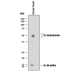
- Detection of Human IL-16 by Western Blot. Western blot shows lysate of human tonsil tissue. PVDF membrane was probed with 1 µg/mL of Goat Anti-Human IL-16 C-terminal Peptide Antigen Affinity-purified Polyclonal Antibody (Catalog # AF-316-PB) followed by HRP-conjugated Anti-Goat IgG Secondary Antibody (Catalog # HAF017). Specific bands were detected for IL-16 at approximately 14 kDa (active) and 45-55 kDa (precursor), as indicated. This experiment was conducted under reducing conditions and using Immunoblot Buffer Group 1.

Supportive validation
- Submitted by
- R&D Systems (provider)
- Main image

- Experimental details
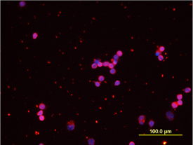
- IL-16 in Human PBMCs. IL-16 was detected in immersion fixed LPS-stimulated human peripheral blood mononuclear cells (PBMCs) using 10 µg/mL Human IL-16 C-terminal Peptide Antigen Affinity-purified Polyclonal Antibody (Catalog # AF-316-PB) for 3 hours at room temperature. Cells were stained with the NorthernLights™ 557-conjugated Anti-Goat IgG Secondary Antibody (red; Catalog # NL001) and counterstained with DAPI (blue). View our protocol for Fluorescent ICC Staining of Non-adherent Cells.
Supportive validation
- Submitted by
- R&D Systems (provider)
- Main image
- Experimental details
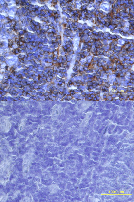
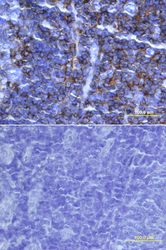
- IL-16 in Human Tonsil. IL-16 was detected in immersion fixed paraffin-embedded sections of human tonsil using 15 µg/mL Human IL-16 Antigen Affinity-purified Polyclonal Antibody (Catalog # AF-316-PB) overnight at 4 °C. Tissue was stained with the Anti-Goat HRP-DAB Cell & Tissue Staining Kit (brown; Catalog # CTS008) and counterstained with hematoxylin (blue). Lower panel shows a lack of labeling if primary antibodies are omitted and tissue is stained only with secondary antibody followed by incubation with detection reagents. View our protocol for Chromogenic IHC Staining of Paraffin-embedded Tissue Sections.