 Explore
 Validate
 Learn
Learn Western blot
 Immunohistochemistry
Antibody data
- Antibody Data
- Antigen structure
- References [7]
- Comments [0]
- Validations
- Immunohistochemistry [2]
- Flow cytometry [1]
- Other assay [5]
Submit
Validation data
Reference
Comment
Report error
- Product number
- 44-550G - Provider product page

- Provider
- Invitrogen Antibodies
- Product name
- Phospho-IRS1 (Ser616) Polyclonal Antibody
- Antibody type
- Polyclonal
- Antigen
- Synthetic peptide
- Description
- Purified from rabbit serum by sequential epitope-specific chromatography, this product contains enough material for 10 mini-blots. This antibody has been negatively preadsorbed using a non-phosphopeptide corresponding to the site of phosphorylation to remove antibody that is reactive with non-phosphorylated insulin receptor substrate-1 (IRS-1). The final product is generated by affinity chromatography using IRS-1 derived peptide that is phosphorylated at serine 616. The antibody has been used in western blotting. Previous lots of this antibody have been used in immunohistochemistry. In western analysis the positive control used was human embryonic kidney cell line transfected with a vector encoding IRS-1 (293T) and stimulated with a phorbol ester, TPA.
- Reactivity
- Human, Mouse, Rat
- Host
- Rabbit
- Isotype
- IgG
- Vial size
- 100 μL
- Storage
- -20°C
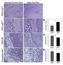
Submitted references Induction of Brain Insulin Resistance and Alzheimer's Molecular Changes by Western Diet.
Altered Insulin Receptor Substrate 1 Phosphorylation in Blood Neuron-Derived Extracellular Vesicles From Patients With Parkinson's Disease.
Central insulin dysregulation and energy dyshomeostasis in two mouse models of Alzheimer's disease.
Insulin and IGF1 signalling pathways in human astrocytes in vitro and in vivo; characterisation, subcellular localisation and modulation of the receptors.
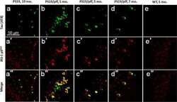
Abnormal serine phosphorylation of insulin receptor substrate 1 is associated with tau pathology in Alzheimer's disease and tauopathies.
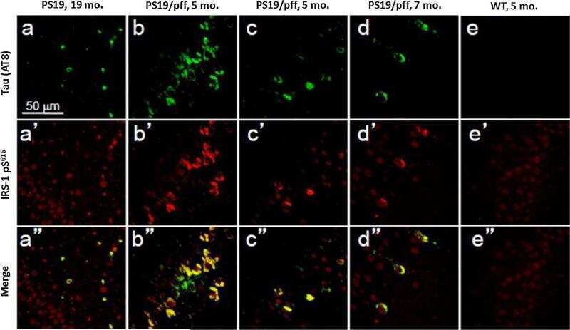
High fat diet produces brain insulin resistance, synaptodendritic abnormalities and altered behavior in mice.
Inhibition of FAK signaling activated by urokinase receptor induces dormancy in human carcinoma cells in vivo.
Mietelska-Porowska A, Domańska J, Want A, Więckowska-Gacek A, Chutorański D, Koperski M, Wojda U
International journal of molecular sciences 2022 Apr 25;23(9)
International journal of molecular sciences 2022 Apr 25;23(9)
Altered Insulin Receptor Substrate 1 Phosphorylation in Blood Neuron-Derived Extracellular Vesicles From Patients With Parkinson's Disease.
Chou SY, Chan L, Chung CC, Chiu JY, Hsieh YC, Hong CT
Frontiers in cell and developmental biology 2020;8:564641
Frontiers in cell and developmental biology 2020;8:564641
Central insulin dysregulation and energy dyshomeostasis in two mouse models of Alzheimer's disease.
Velazquez R, Tran A, Ishimwe E, Denner L, Dave N, Oddo S, Dineley KT
Neurobiology of aging 2017 Oct;58:1-13
Neurobiology of aging 2017 Oct;58:1-13
Insulin and IGF1 signalling pathways in human astrocytes in vitro and in vivo; characterisation, subcellular localisation and modulation of the receptors.
Garwood CJ, Ratcliffe LE, Morgan SV, Simpson JE, Owens H, Vazquez-Villaseñor I, Heath PR, Romero IA, Ince PG, Wharton SB
Molecular brain 2015 Aug 22;8:51
Molecular brain 2015 Aug 22;8:51
Abnormal serine phosphorylation of insulin receptor substrate 1 is associated with tau pathology in Alzheimer's disease and tauopathies.
Yarchoan M, Toledo JB, Lee EB, Arvanitakis Z, Kazi H, Han LY, Louneva N, Lee VM, Kim SF, Trojanowski JQ, Arnold SE
Acta neuropathologica 2014 Nov;128(5):679-89
Acta neuropathologica 2014 Nov;128(5):679-89
High fat diet produces brain insulin resistance, synaptodendritic abnormalities and altered behavior in mice.
Arnold SE, Lucki I, Brookshire BR, Carlson GC, Browne CA, Kazi H, Bang S, Choi BR, Chen Y, McMullen MF, Kim SF
Neurobiology of disease 2014 Jul;67:79-87
Neurobiology of disease 2014 Jul;67:79-87
Inhibition of FAK signaling activated by urokinase receptor induces dormancy in human carcinoma cells in vivo.
Aguirre Ghiso JA
Oncogene 2002 Apr 11;21(16):2513-24
Oncogene 2002 Apr 11;21(16):2513-24
No comments: Submit comment
Supportive validation
- Submitted by
- Invitrogen Antibodies (provider)
- Main image

- Experimental details
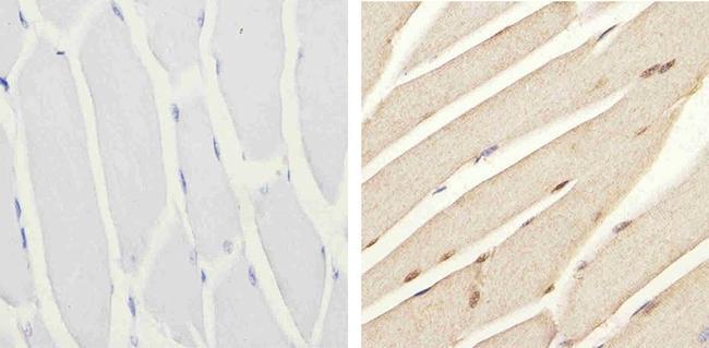
- Immunohistochemistry analysis of Phospho-IRS1 (pS616) showing staining in the nucleus of paraffin-embedded mouse skeletal muscle tissue (right) compared to a negative control without primary antibody (left). To expose target proteins, antigen retrieval was performed using 10mM sodium citrate (pH 6.0), microwaved for 8-15 min. Following antigen retrieval, tissues were blocked in 3% H2O2-methanol for 15 min at room temperature, washed with ddH2O and PBS, and then probed with a Phospho-IRS1 (pS616) polyclonal antibody (Product # 44-550G) diluted in 3% BSA-PBS at a dilution of 1:20 overnight at 4ºC in a humidified chamber. Tissues were washed extensively in PBST and detection was performed using an HRP-conjugated secondary antibody followed by colorimetric detection using a DAB kit. Tissues were counterstained with hematoxylin and dehydrated with ethanol and xylene to prep for mounting.
- Submitted by
- Invitrogen Antibodies (provider)
- Main image

- Experimental details
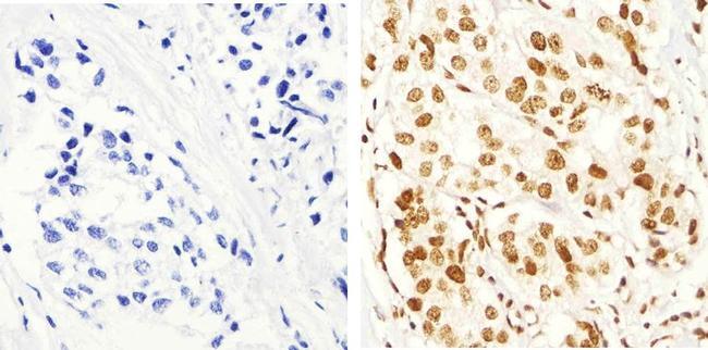
- Immunohistochemistry analysis of Phospho-IRS1 (pS616) showing staining in the nucleus of paraffin-embedded human breast carcinoma tissue (right) compared to a negative control without primary antibody (left). To expose target proteins, antigen retrieval was performed using 10mM sodium citrate (pH 6.0), microwaved for 8-15 min. Following antigen retrieval, tissues were blocked in 3% H2O2-methanol for 15 min at room temperature, washed with ddH2O and PBS, and then probed with a Phospho-IRS1 (pS616) polyclonal antibody (Product # 44-550G) diluted in 3% BSA-PBS at a dilution of 1:100 overnight at 4ºC in a humidified chamber. Tissues were washed extensively in PBST and detection was performed using an HRP-conjugated secondary antibody followed by colorimetric detection using a DAB kit. Tissues were counterstained with hematoxylin and dehydrated with ethanol and xylene to prep for mounting.
Supportive validation
- Submitted by
- Invitrogen Antibodies (provider)
- Main image
- Experimental details
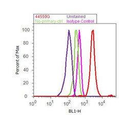
- Flow cytometry analysis of IRS1 [pS616] was done on MCF7 cells treated with Insulin (100nM, 10 minutes). Cells were fixed with 70% ethanol for 10 minutes, permeabilized with 0.25% Triton™ X-100 for 20 minutes, and blocked with 5% BSA for 30 minutes at room temperature. Cells were labeled with IRS1 [pS616] Rabbit Polyclonal Antibody (44550G, red histogram) or with rabbit isotype control (pink histogram) at 3-5 ug/million cells in 2.5% BSA. After incubation at room temperature for 2 hours, the cells were labeled with Alexa Fluor® 488 Goat Anti-Rabbit Secondary Antibody (A11008) at a dilution of 1:400 for 30 minutes at room temperature. The representative 10,000 cells were acquired and analyzed for each sample using an Attune® Acoustic Focusing Cytometer. The purple histogram represents unstained control cells and the green histogram represents no-primary-antibody control.

Supportive validation
- Submitted by
- Invitrogen Antibodies (provider)
- Main image

- Experimental details
- NULL
- Submitted by
- Invitrogen Antibodies (provider)
- Main image
- Experimental details
- NULL
- Submitted by
- Invitrogen Antibodies (provider)
- Main image
- Experimental details
- NULL
- Submitted by
- Invitrogen Antibodies (provider)
- Main image
- Experimental details
- NULL
- Submitted by
- Invitrogen Antibodies (provider)
- Main image
- Experimental details
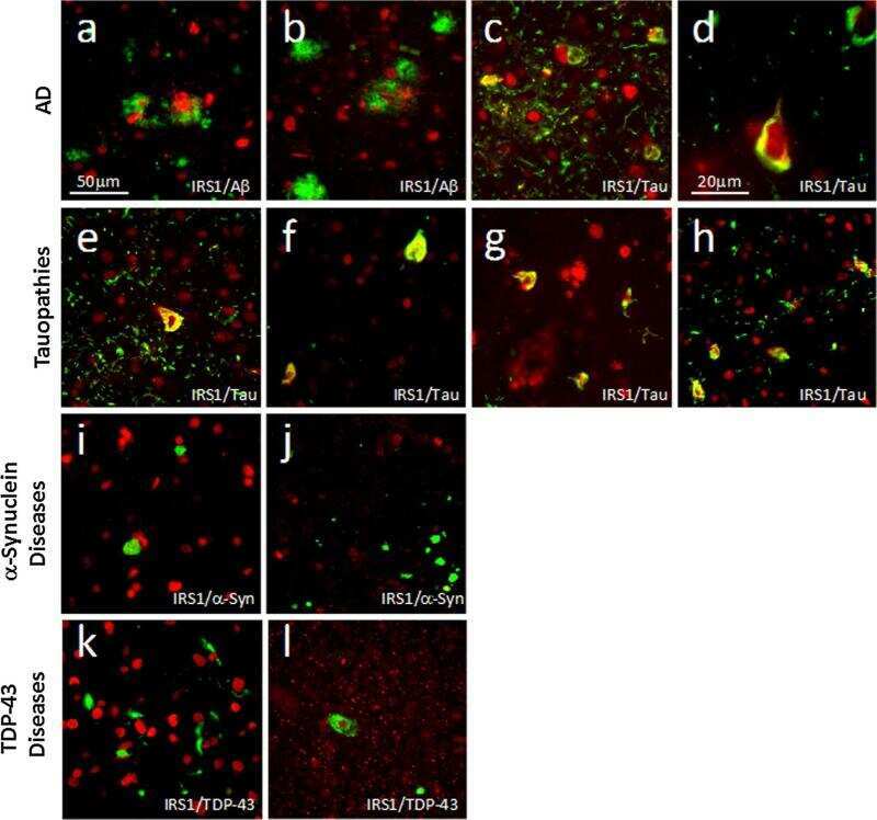
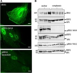
- Fig. 4 Localisation of signalling components in human astrocytes. a Immunocytochemistry demonstrates nuclear localisation of IRS1 phosphorylated at serine 616 (S616) and S636/639 but not total IRS1 b Representative immunoblots of fractionated cell lysates showing the localisation of total IRS1 and IRS1 phosphorylated at S616 and tyrosine 612 (Y612) by immunoblot. Loading control for cytosplasmic fraction are EEF2 and for nuclear fraction is SP1. Molecular weight markers are indicated (kDa)
