Explore
 Validate
 Learn
Learn Western blot
 Immunohistochemistry
Antibody data
- Antibody Data
- Antigen structure
- References [46]
- Comments [0]
- Validations
- Immunohistochemistry [1]
- Flow cytometry [1]
- Other assay [23]
Submit
Validation data
Reference
Comment
Report error
- Product number
- 44-816G - Provider product page

- Provider
- Invitrogen Antibodies
- Product name
- Phospho-IRS1 (Tyr612) Polyclonal Antibody
- Antibody type
- Polyclonal
- Antigen
- Synthetic peptide
- Description
- On western blotting this antibody detects a protein with the apparent MW of 165 kDa. Positive controls used were Insulin (50 nM for 5 minutes) or TPA (100 ng/mL for 30 min) treated human embryonic kidney cells (293T) or Chinese Hamster Ovary cells (CHO-T) transiently transfected with a plasmid encoding human IRS1.
- Reactivity
- Human
- Host
- Rabbit
- Isotype
- IgG
- Vial size
- 100 μL
- Storage
- -20°C
Submitted references Repurposing Ceritinib Induces DNA Damage and Enhances PARP Inhibitor Responses in High-Grade Serous Ovarian Carcinoma.
A Phase 0 Trial of Ceritinib in Patients with Brain Metastases and Recurrent Glioblastoma.
Cullin neddylation inhibitor attenuates hyperglycemia by enhancing hepatic insulin signaling through insulin receptor substrate stabilization.
Exercise Counterbalances Rho/ROCK2 Signaling Impairment in the Skeletal Muscle and Ameliorates Insulin Sensitivity in Obese Mice.
Insulin Resistance and Pellino-1 Mediated Decrease in the Activities of Vasodilator Signaling Contributes to Sunitinib-Induced Hypertension.
JMJD8 is a Novel Molecular Nexus Between Adipocyte-Intrinsic Inflammation and Insulin Resistance.
The autophagy protein Becn1 improves insulin sensitivity by promoting adiponectin secretion via exocyst binding.
The anti-diabetic effects of NAG-1/GDF15 on HFD/STZ-induced mice.
The E3 ligase TRAF4 promotes IGF signaling by mediating atypical ubiquitination of IRS-1.
Role of Exendin-4 in Brain Insulin Resistance, Mitochondrial Function, and Neurite Outgrowth in Neurons under Palmitic Acid-Induced Oxidative Stress.
Altered Insulin Receptor Substrate 1 Phosphorylation in Blood Neuron-Derived Extracellular Vesicles From Patients With Parkinson's Disease.
Direct Effects of D-Chiro-Inositol on Insulin Signaling and Glucagon Secretion of Pancreatic Alpha Cells.
Intranasal insulin administration may be highly effective in improving cognitive function in mice with cognitive dysfunction by reversing brain insulin resistance.
IPF pathogenesis is dependent upon TGFβ induction of IGF-1.
Iron overload inhibits late stage autophagic flux leading to insulin resistance.
The Diurnal Rhythm of Insulin Receptor Substrate-1 (IRS-1) and Kir4.1 in Diabetes: Implications for a Clock Gene Bmal1.
Coenzyme Q10 protects against burn-induced mitochondrial dysfunction and impaired insulin signaling in mouse skeletal muscle.
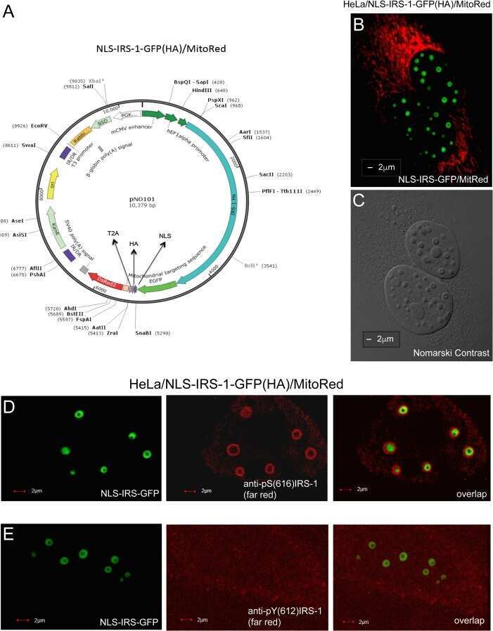
Molecular and Structural Traits of Insulin Receptor Substrate 1/LC3 Nuclear Structures and Their Role in Autophagy Control and Tumor Cell Survival.
Ablation of Grb10 Specifically in Muscle Impacts Muscle Size and Glucose Metabolism in Mice.
Chronic restraint stress induces hippocampal memory deficits by impairing insulin signaling.
TET2 facilitates PPARγ agonist-mediated gene regulation and insulin sensitization in adipocytes.
Dnmt3a is an epigenetic mediator of adipose insulin resistance.
Tau deletion promotes brain insulin resistance.
Epigenetic regulation of macrophage polarization and inflammation by DNA methylation in obesity.
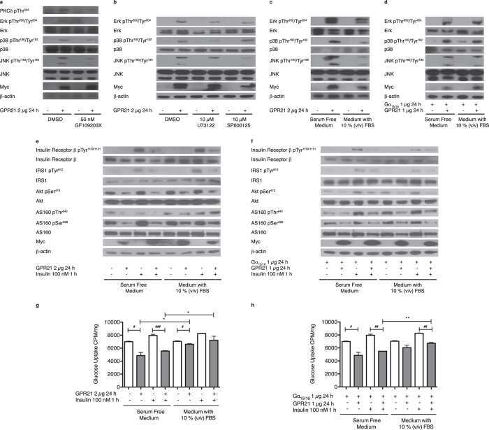
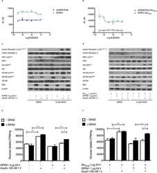
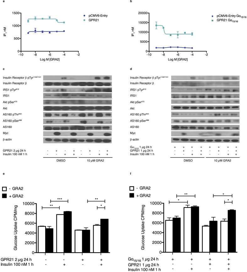
Regulating the effects of GPR21, a novel target for type 2 diabetes.
Role of protein farnesylation in burn-induced metabolic derangements and insulin resistance in mouse skeletal muscle.
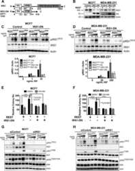
Type 1 Insulin-Like Growth Factor Receptor/Insulin Receptor Substrate 1 Signaling Confers Pathogenic Activity on Breast Tumor Cells Lacking REST.
Severe insulin resistance alters metabolism in mesenchymal progenitor cells.
Basal expression of insulin-like growth factor 1 receptor determines intrinsic resistance of cancer cells to a phosphatidylinositol 3-kinase inhibitor ZSTK474.
Fetuin B Is a Secreted Hepatocyte Factor Linking Steatosis to Impaired Glucose Metabolism.
Insulin and IGF1 signalling pathways in human astrocytes in vitro and in vivo; characterisation, subcellular localisation and modulation of the receptors.
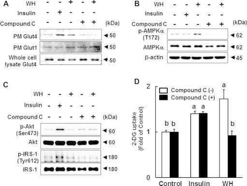
The di-peptide Trp-His activates AMP-activated protein kinase and enhances glucose uptake independently of insulin in L6 myotubes.
Salicylate acutely stimulates 5'-AMP-activated protein kinase and insulin-independent glucose transport in rat skeletal muscles.
PTEN is a protein tyrosine phosphatase for IRS1.
A novel inhibitor of the insulin/IGF signaling pathway protects from age-onset, neurodegeneration-linked proteotoxicity.
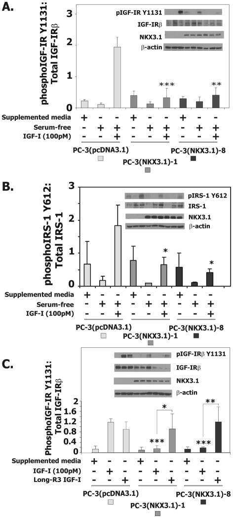
Variant NKX3.1 and Serum IGF-1: Investigation of Interaction in Prostate Cancer.
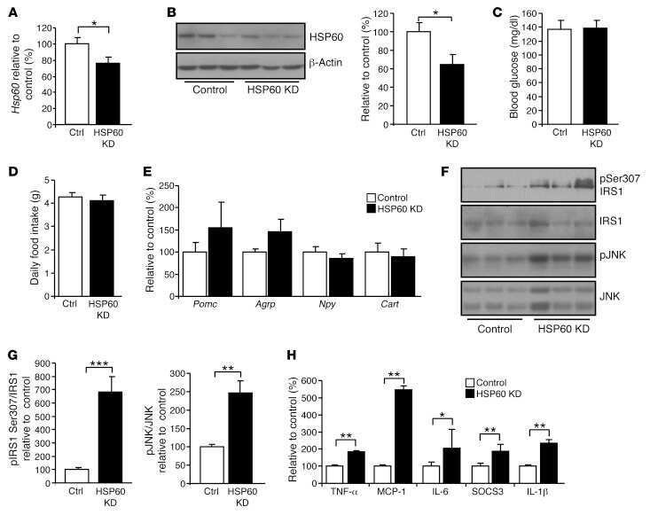
Leptin regulation of Hsp60 impacts hypothalamic insulin signaling.
Resistance exercise, but not endurance exercise, induces IKKβ phosphorylation in human skeletal muscle of training-accustomed individuals.
C1q/TNF-related protein-12 (CTRP12), a novel adipokine that improves insulin sensitivity and glycemic control in mouse models of obesity and diabetes.
Obesity in mice with adipocyte-specific deletion of clock component Arntl.
Role of TAPP1 and TAPP2 adaptor binding to PtdIns(3,4)P2 in regulating insulin sensitivity defined by knock-in analysis.
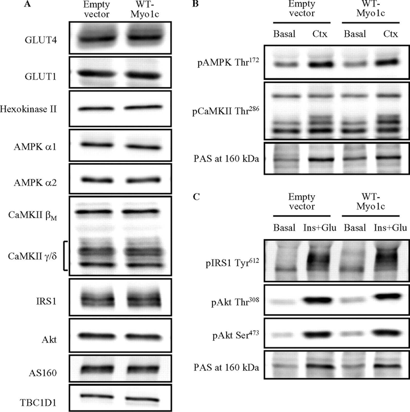
Myo1c regulates glucose uptake in mouse skeletal muscle.
High IGF-IR activity in triple-negative breast cancer cell lines and tumorgrafts correlates with sensitivity to anti-IGF-IR therapy.
Compensatory insulin receptor (IR) activation on inhibition of insulin-like growth factor-1 receptor (IGF-1R): rationale for cotargeting IGF-1R and IR in cancer.
NKX3.1 activates expression of insulin-like growth factor binding protein-3 to mediate insulin-like growth factor-I signaling and cell proliferation.
Insulin-like growth factor-I activation of Akt survival cascade in neuronal cells requires the presence of its cognate receptor in caveolae.
Kanakkanthara A, Hou X, Ekstrom TL, Zanfagnin V, Huehls AM, Kelly RL, Ding H, Larson MC, Vasmatzis G, Oberg AL, Kaufmann SH, Mansfield AS, Weroha SJ, Karnitz LM
Cancer research 2022 Jan 15;82(2):307-319
Cancer research 2022 Jan 15;82(2):307-319
A Phase 0 Trial of Ceritinib in Patients with Brain Metastases and Recurrent Glioblastoma.
Mehta S, Fiorelli R, Bao X, Pennington-Krygier C, Derogatis A, Kim S, Yoo W, Li J, Sanai N
Clinical cancer research : an official journal of the American Association for Cancer Research 2022 Jan 15;28(2):289-297
Clinical cancer research : an official journal of the American Association for Cancer Research 2022 Jan 15;28(2):289-297
Cullin neddylation inhibitor attenuates hyperglycemia by enhancing hepatic insulin signaling through insulin receptor substrate stabilization.
Chen C, Gu L, Matye DJ, Clayton YD, Hasan MN, Wang Y, Friedman JE, Li T
Proceedings of the National Academy of Sciences of the United States of America 2022 Feb 8;119(6)
Proceedings of the National Academy of Sciences of the United States of America 2022 Feb 8;119(6)
Exercise Counterbalances Rho/ROCK2 Signaling Impairment in the Skeletal Muscle and Ameliorates Insulin Sensitivity in Obese Mice.
Muñoz VR, Gaspar RC, Severino MB, Macêdo APA, Simabuco FM, Ropelle ER, Cintra DE, da Silva ASR, Kim YB, Pauli JR
Frontiers in immunology 2021;12:702025
Frontiers in immunology 2021;12:702025
Insulin Resistance and Pellino-1 Mediated Decrease in the Activities of Vasodilator Signaling Contributes to Sunitinib-Induced Hypertension.
Liu Y, Tang LL, Liang C, Wu MM, Zhang ZR
Frontiers in pharmacology 2021;12:617165
Frontiers in pharmacology 2021;12:617165
JMJD8 is a Novel Molecular Nexus Between Adipocyte-Intrinsic Inflammation and Insulin Resistance.
You D, Chul Jung B, Villivalam SD, Lim HW, Kang S
Diabetes 2021 Oct 21;
Diabetes 2021 Oct 21;
The autophagy protein Becn1 improves insulin sensitivity by promoting adiponectin secretion via exocyst binding.
Kuramoto K, Kim YJ, Hong JH, He C
Cell reports 2021 May 25;35(8):109184
Cell reports 2021 May 25;35(8):109184
The anti-diabetic effects of NAG-1/GDF15 on HFD/STZ-induced mice.
Lertpatipanpong P, Lee J, Kim I, Eling T, Oh SY, Seong JK, Baek SJ
Scientific reports 2021 Jul 22;11(1):15027
Scientific reports 2021 Jul 22;11(1):15027
The E3 ligase TRAF4 promotes IGF signaling by mediating atypical ubiquitination of IRS-1.
Yu W, Singh R, Wang Z, O'Malley BW, Yi P
The Journal of biological chemistry 2021 Jan-Jun;296:100739
The Journal of biological chemistry 2021 Jan-Jun;296:100739
Role of Exendin-4 in Brain Insulin Resistance, Mitochondrial Function, and Neurite Outgrowth in Neurons under Palmitic Acid-Induced Oxidative Stress.
Jo D, Yoon G, Song J
Antioxidants (Basel, Switzerland) 2021 Jan 9;10(1)
Antioxidants (Basel, Switzerland) 2021 Jan 9;10(1)
Altered Insulin Receptor Substrate 1 Phosphorylation in Blood Neuron-Derived Extracellular Vesicles From Patients With Parkinson's Disease.
Chou SY, Chan L, Chung CC, Chiu JY, Hsieh YC, Hong CT
Frontiers in cell and developmental biology 2020;8:564641
Frontiers in cell and developmental biology 2020;8:564641
Direct Effects of D-Chiro-Inositol on Insulin Signaling and Glucagon Secretion of Pancreatic Alpha Cells.
Filippello A, Scamporrino A, Di Mauro S, Malaguarnera R, Di Pino A, Scicali R, Purrello F, Piro S
Biomolecules 2020 Oct 4;10(10)
Biomolecules 2020 Oct 4;10(10)
Intranasal insulin administration may be highly effective in improving cognitive function in mice with cognitive dysfunction by reversing brain insulin resistance.
Lv H, Tang L, Guo C, Jiang Y, Gao C, Wang Y, Jian C
Cognitive neurodynamics 2020 Jun;14(3):323-338
Cognitive neurodynamics 2020 Jun;14(3):323-338
IPF pathogenesis is dependent upon TGFβ induction of IGF-1.
Hernandez DM, Kang JH, Choudhury M, Andrianifahanana M, Yin X, Limper AH, Leof EB
FASEB journal : official publication of the Federation of American Societies for Experimental Biology 2020 Apr;34(4):5363-5388
FASEB journal : official publication of the Federation of American Societies for Experimental Biology 2020 Apr;34(4):5363-5388
Iron overload inhibits late stage autophagic flux leading to insulin resistance.
Jahng JWS, Alsaadi RM, Palanivel R, Song E, Hipolito VEB, Sung HK, Botelho RJ, Russell RC, Sweeney G
EMBO reports 2019 Oct 4;20(10):e47911
EMBO reports 2019 Oct 4;20(10):e47911
The Diurnal Rhythm of Insulin Receptor Substrate-1 (IRS-1) and Kir4.1 in Diabetes: Implications for a Clock Gene Bmal1.
Luo Q, Xiao Y, Alex A, Cummins TR, Bhatwadekar AD
Investigative ophthalmology & visual science 2019 May 1;60(6):1928-1936
Investigative ophthalmology & visual science 2019 May 1;60(6):1928-1936
Coenzyme Q10 protects against burn-induced mitochondrial dysfunction and impaired insulin signaling in mouse skeletal muscle.
Nakazawa H, Ikeda K, Shinozaki S, Yasuhara S, Yu YM, Martyn JAJ, Tompkins RG, Yorozu T, Inoue S, Kaneki M
FEBS open bio 2019 Feb;9(2):348-363
FEBS open bio 2019 Feb;9(2):348-363
Molecular and Structural Traits of Insulin Receptor Substrate 1/LC3 Nuclear Structures and Their Role in Autophagy Control and Tumor Cell Survival.
Lassak A, Dean M, Wyczechowska D, Wilk A, Marrero L, Trillo-Tinoco J, Boulares AH, Sarkaria JN, Del Valle L, Peruzzi F, Ochoa A, Reiss K
Molecular and cellular biology 2018 May 15;38(10)
Molecular and cellular biology 2018 May 15;38(10)
Ablation of Grb10 Specifically in Muscle Impacts Muscle Size and Glucose Metabolism in Mice.
Holt LJ, Brandon AE, Small L, Suryana E, Preston E, Wilks D, Mokbel N, Coles CA, White JD, Turner N, Daly RJ, Cooney GJ
Endocrinology 2018 Mar 1;159(3):1339-1351
Endocrinology 2018 Mar 1;159(3):1339-1351
Chronic restraint stress induces hippocampal memory deficits by impairing insulin signaling.
Woo H, Hong CJ, Jung S, Choe S, Yu SW
Molecular brain 2018 Jul 3;11(1):37
Molecular brain 2018 Jul 3;11(1):37
TET2 facilitates PPARγ agonist-mediated gene regulation and insulin sensitization in adipocytes.
Bian F, Ma X, Villivalam SD, You D, Choy LR, Paladugu A, Fung S, Kang S
Metabolism: clinical and experimental 2018 Dec;89:39-47
Metabolism: clinical and experimental 2018 Dec;89:39-47
Dnmt3a is an epigenetic mediator of adipose insulin resistance.
You D, Nilsson E, Tenen DE, Lyubetskaya A, Lo JC, Jiang R, Deng J, Dawes BA, Vaag A, Ling C, Rosen ED, Kang S
eLife 2017 Nov 1;6
eLife 2017 Nov 1;6
Tau deletion promotes brain insulin resistance.
Marciniak E, Leboucher A, Caron E, Ahmed T, Tailleux A, Dumont J, Issad T, Gerhardt E, Pagesy P, Vileno M, Bournonville C, Hamdane M, Bantubungi K, Lancel S, Demeyer D, Eddarkaoui S, Vallez E, Vieau D, Humez S, Faivre E, Grenier-Boley B, Outeiro TF, Staels B, Amouyel P, Balschun D, Buee L, Blum D
The Journal of experimental medicine 2017 Aug 7;214(8):2257-2269
The Journal of experimental medicine 2017 Aug 7;214(8):2257-2269
Epigenetic regulation of macrophage polarization and inflammation by DNA methylation in obesity.
Wang X, Cao Q, Yu L, Shi H, Xue B, Shi H
JCI insight 2016 Nov 17;1(19):e87748
JCI insight 2016 Nov 17;1(19):e87748
Regulating the effects of GPR21, a novel target for type 2 diabetes.
Leonard S, Kinsella GK, Benetti E, Findlay JBC
Scientific reports 2016 May 31;6:27002
Scientific reports 2016 May 31;6:27002
Role of protein farnesylation in burn-induced metabolic derangements and insulin resistance in mouse skeletal muscle.
Nakazawa H, Yamada M, Tanaka T, Kramer J, Yu YM, Fischman AJ, Martyn JA, Tompkins RG, Kaneki M
PloS one 2015;10(1):e0116633
PloS one 2015;10(1):e0116633
Type 1 Insulin-Like Growth Factor Receptor/Insulin Receptor Substrate 1 Signaling Confers Pathogenic Activity on Breast Tumor Cells Lacking REST.
Meyer K, Albaugh B, Schoenike B, Roopra A
Molecular and cellular biology 2015 Sep 1;35(17):2991-3004
Molecular and cellular biology 2015 Sep 1;35(17):2991-3004
Severe insulin resistance alters metabolism in mesenchymal progenitor cells.
Balhara B, Burkart A, Topcu V, Lee YK, Cowan C, Kahn CR, Patti ME
Endocrinology 2015 Jun;156(6):2039-48
Endocrinology 2015 Jun;156(6):2039-48
Basal expression of insulin-like growth factor 1 receptor determines intrinsic resistance of cancer cells to a phosphatidylinositol 3-kinase inhibitor ZSTK474.
Isoyama S, Kajiwara G, Tamaki N, Okamura M, Yoshimi H, Nakamura N, Kawamura K, Nishimura Y, Namatame N, Yamori T, Dan S
Cancer science 2015 Feb;106(2):171-8
Cancer science 2015 Feb;106(2):171-8
Fetuin B Is a Secreted Hepatocyte Factor Linking Steatosis to Impaired Glucose Metabolism.
Meex RC, Hoy AJ, Morris A, Brown RD, Lo JC, Burke M, Goode RJ, Kingwell BA, Kraakman MJ, Febbraio MA, Greve JW, Rensen SS, Molloy MP, Lancaster GI, Bruce CR, Watt MJ
Cell metabolism 2015 Dec 1;22(6):1078-89
Cell metabolism 2015 Dec 1;22(6):1078-89
Insulin and IGF1 signalling pathways in human astrocytes in vitro and in vivo; characterisation, subcellular localisation and modulation of the receptors.
Garwood CJ, Ratcliffe LE, Morgan SV, Simpson JE, Owens H, Vazquez-Villaseñor I, Heath PR, Romero IA, Ince PG, Wharton SB
Molecular brain 2015 Aug 22;8:51
Molecular brain 2015 Aug 22;8:51
The di-peptide Trp-His activates AMP-activated protein kinase and enhances glucose uptake independently of insulin in L6 myotubes.
Soga M, Ohashi A, Taniguchi M, Matsui T, Tsuda T
FEBS open bio 2014;4:898-904
FEBS open bio 2014;4:898-904
Salicylate acutely stimulates 5'-AMP-activated protein kinase and insulin-independent glucose transport in rat skeletal muscles.
Serizawa Y, Oshima R, Yoshida M, Sakon I, Kitani K, Goto A, Tsuda S, Hayashi T
Biochemical and biophysical research communications 2014 Oct 10;453(1):81-5
Biochemical and biophysical research communications 2014 Oct 10;453(1):81-5
PTEN is a protein tyrosine phosphatase for IRS1.
Shi Y, Wang J, Chandarlapaty S, Cross J, Thompson C, Rosen N, Jiang X
Nature structural & molecular biology 2014 Jun;21(6):522-7
Nature structural & molecular biology 2014 Jun;21(6):522-7
A novel inhibitor of the insulin/IGF signaling pathway protects from age-onset, neurodegeneration-linked proteotoxicity.
El-Ami T, Moll L, Carvalhal Marques F, Volovik Y, Reuveni H, Cohen E
Aging cell 2014 Feb;13(1):165-74
Aging cell 2014 Feb;13(1):165-74
Variant NKX3.1 and Serum IGF-1: Investigation of Interaction in Prostate Cancer.
Muhlbradt E, Ma J, Severi G, Ortner E, Hayes V, Hoang HN, Stampfer M, Giles G, Pollak M, Gelmann EP
Genes & cancer 2013 Nov;4(11-12):535-45
Genes & cancer 2013 Nov;4(11-12):535-45
Leptin regulation of Hsp60 impacts hypothalamic insulin signaling.
Kleinridders A, Lauritzen HP, Ussar S, Christensen JH, Mori MA, Bross P, Kahn CR
The Journal of clinical investigation 2013 Nov;123(11):4667-80
The Journal of clinical investigation 2013 Nov;123(11):4667-80
Resistance exercise, but not endurance exercise, induces IKKβ phosphorylation in human skeletal muscle of training-accustomed individuals.
Møller AB, Vendelbo MH, Rahbek SK, Clasen BF, Schjerling P, Vissing K, Jessen N
Pflugers Archiv : European journal of physiology 2013 Dec;465(12):1785-95
Pflugers Archiv : European journal of physiology 2013 Dec;465(12):1785-95
C1q/TNF-related protein-12 (CTRP12), a novel adipokine that improves insulin sensitivity and glycemic control in mouse models of obesity and diabetes.
Wei Z, Peterson JM, Lei X, Cebotaru L, Wolfgang MJ, Baldeviano GC, Wong GW
The Journal of biological chemistry 2012 Mar 23;287(13):10301-10315
The Journal of biological chemistry 2012 Mar 23;287(13):10301-10315
Obesity in mice with adipocyte-specific deletion of clock component Arntl.
Paschos GK, Ibrahim S, Song WL, Kunieda T, Grant G, Reyes TM, Bradfield CA, Vaughan CH, Eiden M, Masoodi M, Griffin JL, Wang F, Lawson JA, Fitzgerald GA
Nature medicine 2012 Dec;18(12):1768-77
Nature medicine 2012 Dec;18(12):1768-77
Role of TAPP1 and TAPP2 adaptor binding to PtdIns(3,4)P2 in regulating insulin sensitivity defined by knock-in analysis.
Wullschleger S, Wasserman DH, Gray A, Sakamoto K, Alessi DR
The Biochemical journal 2011 Mar 1;434(2):265-74
The Biochemical journal 2011 Mar 1;434(2):265-74
Myo1c regulates glucose uptake in mouse skeletal muscle.
Toyoda T, An D, Witczak CA, Koh HJ, Hirshman MF, Fujii N, Goodyear LJ
The Journal of biological chemistry 2011 Feb 11;286(6):4133-40
The Journal of biological chemistry 2011 Feb 11;286(6):4133-40
High IGF-IR activity in triple-negative breast cancer cell lines and tumorgrafts correlates with sensitivity to anti-IGF-IR therapy.
Litzenburger BC, Creighton CJ, Tsimelzon A, Chan BT, Hilsenbeck SG, Wang T, Carboni JM, Gottardis MM, Huang F, Chang JC, Lewis MT, Rimawi MF, Lee AV
Clinical cancer research : an official journal of the American Association for Cancer Research 2011 Apr 15;17(8):2314-27
Clinical cancer research : an official journal of the American Association for Cancer Research 2011 Apr 15;17(8):2314-27
Compensatory insulin receptor (IR) activation on inhibition of insulin-like growth factor-1 receptor (IGF-1R): rationale for cotargeting IGF-1R and IR in cancer.
Buck E, Gokhale PC, Koujak S, Brown E, Eyzaguirre A, Tao N, Rosenfeld-Franklin M, Lerner L, Chiu MI, Wild R, Epstein D, Pachter JA, Miglarese MR
Molecular cancer therapeutics 2010 Oct;9(10):2652-64
Molecular cancer therapeutics 2010 Oct;9(10):2652-64
NKX3.1 activates expression of insulin-like growth factor binding protein-3 to mediate insulin-like growth factor-I signaling and cell proliferation.
Muhlbradt E, Asatiani E, Ortner E, Wang A, Gelmann EP
Cancer research 2009 Mar 15;69(6):2615-22
Cancer research 2009 Mar 15;69(6):2615-22
Insulin-like growth factor-I activation of Akt survival cascade in neuronal cells requires the presence of its cognate receptor in caveolae.
Lu X, Kambe F, Cao X, Yamauchi M, Seo H
Experimental cell research 2008 Jan 15;314(2):342-51
Experimental cell research 2008 Jan 15;314(2):342-51
No comments: Submit comment
Supportive validation
- Submitted by
- Invitrogen Antibodies (provider)
- Main image

- Experimental details
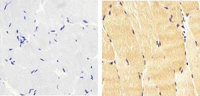
- Immunohistochemistry analysis of Phospho-IRS1 (pY612) showing staining in the cytoplasm and nucleus of paraffin-embedded human skeletal muscle tissue (right) compared to a negative control without primary antibody (left). To expose target proteins, antigen retrieval was performed using 10mM sodium citrate (pH 6.0), microwaved for 8-15 min. Following antigen retrieval, tissues were blocked in 3% H2O2-methanol for 15 min at room temperature, washed with ddH2O and PBS, and then probed with a Phospho-IRS1 (pY612) polyclonal antibody (Product # 44-816G) diluted in 3% BSA-PBS at a dilution of 1:20 overnight at 4ºC in a humidified chamber. Tissues were washed extensively in PBST and detection was performed using an HRP-conjugated secondary antibody followed by colorimetric detection using a DAB kit. Tissues were counterstained with hematoxylin and dehydrated with ethanol and xylene to prep for mounting.
Supportive validation
- Submitted by
- Invitrogen Antibodies (provider)
- Main image
- Experimental details
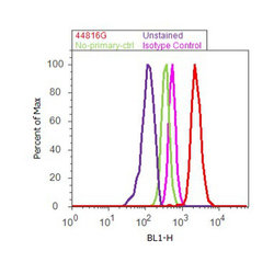
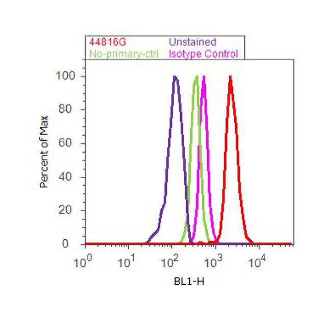
- Flow cytometry analysis of IRS1 [pY612] was done on MCF7 cells treated with Insulin (100nM, 10 minutes). Cells were fixed with 70% ethanol for 10 minutes, permeabilized with 0.25% Triton™ X-100 for 20 minutes, and blocked with 5% BSA for 30 minutes at room temperature. Cells were labeled with IRS1 [pY612] Rabbit Polyclonal Antibody (44816G, red histogram) or with rabbit isotype control (pink histogram) at 3-5 ug/million cells in 2.5% BSA. After incubation at room temperature for 2 hours, the cells were labeled with Alexa Fluor® 488 Goat Anti-Rabbit Secondary Antibody (A11008) at a dilution of 1:400 for 30 minutes at room temperature. The representative 10,000 cells were acquired and analyzed for each sample using an Attune® Acoustic Focusing Cytometer. The purple histogram represents unstained control cells and the green histogram represents no-primary-antibody control.
Supportive validation
- Submitted by
- Invitrogen Antibodies (provider)
- Main image

- Experimental details
- NULL
- Submitted by
- Invitrogen Antibodies (provider)
- Main image
- Experimental details
- NULL

- Submitted by
- Invitrogen Antibodies (provider)
- Main image
- Experimental details
- NULL

- Submitted by
- Invitrogen Antibodies (provider)
- Main image

- Experimental details
- NULL
- Submitted by
- Invitrogen Antibodies (provider)
- Main image
- Experimental details
- NULL

- Submitted by
- Invitrogen Antibodies (provider)
- Main image
- Experimental details
- NULL
- Submitted by
- Invitrogen Antibodies (provider)
- Main image
- Experimental details
- NULL
- Submitted by
- Invitrogen Antibodies (provider)
- Main image
- Experimental details
- NULL
- Submitted by
- Invitrogen Antibodies (provider)
- Main image

- Experimental details
- NULL
- Submitted by
- Invitrogen Antibodies (provider)
- Main image

- Experimental details
- NULL
- Submitted by
- Invitrogen Antibodies (provider)
- Main image

- Experimental details
- NULL
- Submitted by
- Invitrogen Antibodies (provider)
- Main image
- Experimental details
- NULL

- Submitted by
- Invitrogen Antibodies (provider)
- Main image
- Experimental details
- NULL

- Submitted by
- Invitrogen Antibodies (provider)
- Main image
- Experimental details
- NULL

- Submitted by
- Invitrogen Antibodies (provider)
- Main image
- Experimental details
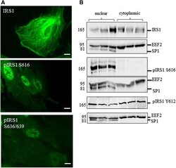
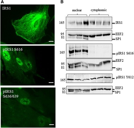
- Fig. 4 Localisation of signalling components in human astrocytes. a Immunocytochemistry demonstrates nuclear localisation of IRS1 phosphorylated at serine 616 (S616) and S636/639 but not total IRS1 b Representative immunoblots of fractionated cell lysates showing the localisation of total IRS1 and IRS1 phosphorylated at S616 and tyrosine 612 (Y612) by immunoblot. Loading control for cytosplasmic fraction are EEF2 and for nuclear fraction is SP1. Molecular weight markers are indicated (kDa)
- Submitted by
- Invitrogen Antibodies (provider)
- Main image
- Experimental details
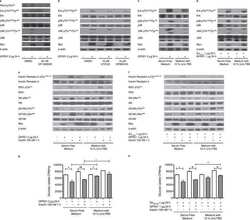
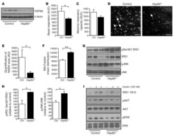
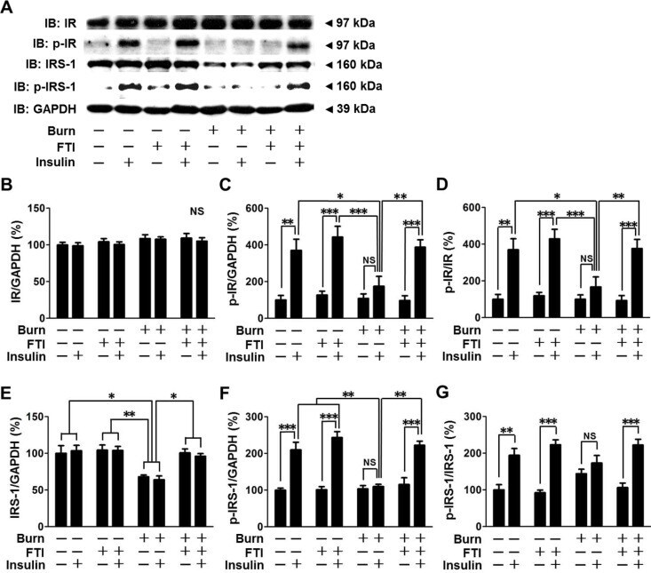
- Figure 2 FTI-277 treatment reversed burn-induced impaired insulin receptor (IR)-insulin receptor substrate-1 (IRS-1) signaling in skeletal muscle. IR protein expression was not altered by burn, FTI-277 or insulin (B). Insulin-stimulated IR phosphorylation was attenuated at 3 days after burn compared with sham-burned mice, which was almost completely restored by FTI-277 treatment (C, D). Burn decreased IRS-1 protein expression (E) and insulin-stimulated IRS-1 phosphorylation (F, G), both of which were reversed by FTI-277. Insulin significantly increased p-IRS-1/GAPDH ratio and p-IRS-1/IRS-1 ratio in sham-burned mice and FTI-277-treated burned mice, whereas insulin failed to significantly increase p-IRS-1/GAPDH ratio or p-IRS-1/IRS-1 ratio in vehicle-treated burned mice (F, G). There was a trend of increase in insulin-stimulated p-IRS-1/IRS-1 ratio in vehicle-treated burned mice, but there was no statistical difference (G). n = 5 mice per group for saline-injected mice, n = 6 mice per group for insulin-injected sham-burned mice, n = 8 mice per group for insulin-injected burned mice. *p
- Submitted by
- Invitrogen Antibodies (provider)
- Main image

- Experimental details
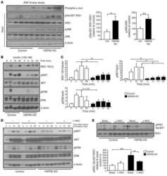
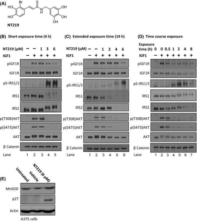
- Figure 1 NT219 inhibits the IGF1R to AKT signaling pathway in human melanoma cells. (A) Chemical formula of NT219. (B-C) NT219 treatment reduces the IGF-induced autophosphorylation of the IGF1R, induces inhibitory Ser-phosphorylation and subsequent elimination of the insulin receptor substrate 1 (IRS1) and 2 (IRS2), and prevents the IGF1-induced activation of AKT in A375 human melanoma cells, following short (B) and long (C) exposures. (D) Temporal analysis of the effects of NT219 on components of the IGF pathway indicates that the compound acts on the IGF1R and AKT within 1 h to block the signaling cascade and confers Ser-phosphorylation and degradation of IRS1 and IRS2 2-8 h after exposure to achieve long-term inhibition. (E) NT219 elevates the expression levels of the FOXO target genes p27 and manganese superoxide dismutase (MnSOD) in A375 cells.
- Submitted by
- Invitrogen Antibodies (provider)
- Main image
- Experimental details
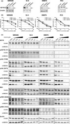
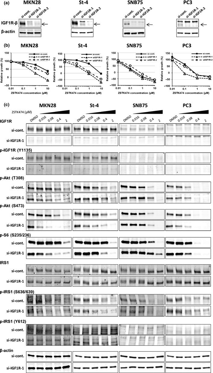
- Fig 3 Effect of insulin-like growth factor 1 receptor (IGF1R) siRNAs on activity of ZSTK474 to inhibit cell proliferation and phosphatidylinositol 3-kinase downstream signaling. (a) Knockdown of IGF1R protein expression using IGF1R siRNAs. MKN28, St-4, SNB75, and PC3 cells were transfected with control siRNA (si-cont.) or IGF1R siRNAs (siIGF1R-1 or siIGF1R-2) and the expression of IGF1R protein was measured by immunoblot analysis. (b) Dose-response curves of ZSTK474 against cell growth in MKN28, St-4, SNB75, and PC3 cells after transfection with control siRNA or IGF1R siRNAs. Cell growth was assessed by sulforhodamine B assay. Assays were carried out in duplicate and the data are representative of two independent experiments. (c) MKN28, St-4, SNB75, and PC3 cells were transfected with either control siRNA or IGF1R siRNA, and then exposed to ZSTK474 at the indicated concentrations for 3 h. Cells were then harvested and immunoblot analyses of indicated proteins were carried out.
- Submitted by
- Invitrogen Antibodies (provider)
- Main image
- Experimental details
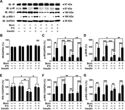
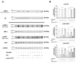
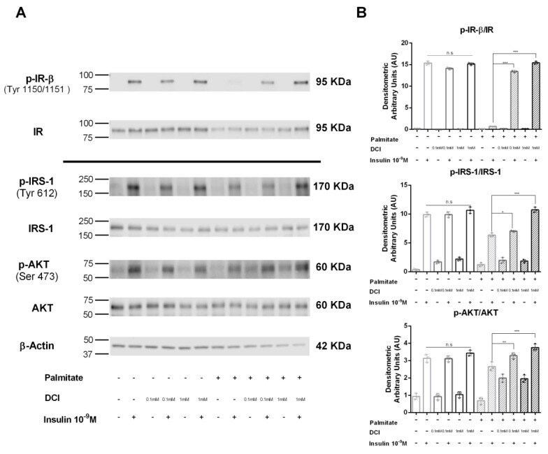
- Figure 5 Effect of DCI on insulin receptor (IR) phosphorylation and the IRS-1/AKT pathway in alpha-TC1-6 cells exposed to palmitate. Immunoprecipitation and Western blot analysis for total IR (Tyr1150/1151-beta subunit) (p-IR-beta) and total IR; Western blot analysis for p-IRS-1 (Tyr612), total IRS-1 (IRS-1), p-AKT (Ser 473), total AKT (AKT), and beta-actin: alphaTC1-6 cells were treated with DCI (0.1 or 1 mM for 48 h) in the presence or absence of palmitate (0.5 mM for 48 h) and acutely stimulated with insulin (10 -9 M for 5 min). ( A ) Representative immunoblot of three independent experiments. ( B ) Means +- SEM of densitometric analysis. One-way ANOVA followed by Bonferroni test ( n = 3) was used to evaluate statistical significance in insulin-stimulated cells treated with increasing concentrations of DCI with respect to insulin-stimulated controls in the absence and presence of palmitate: * p < 0.05, ** p < 0.01, *** p < 0.001. n.s., not significant; DCI, D-chiro-inositol.
- Submitted by
- Invitrogen Antibodies (provider)
- Main image

- Experimental details
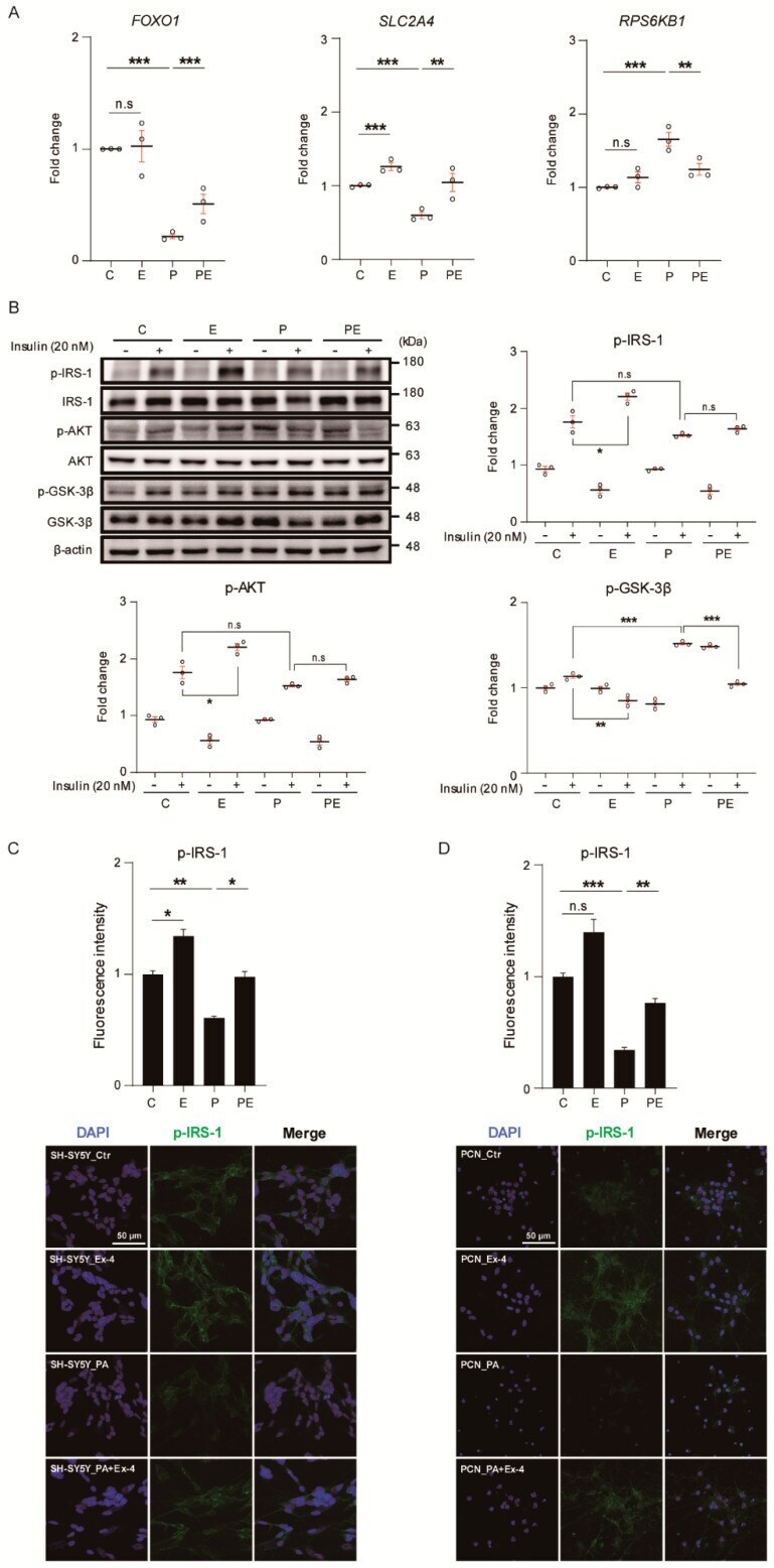
- Figure 2 Exendin-4 improves insulin signaling in a neuron under palmitic acid-induced oxidative stress. ( A ) Comparison of mRNA expression levels of insulin signaling related genes ( FOXO1 , SLC2A4 , and RPS6KB1 ) in SH-SY5Y cells treated with vehicle (C or Ctr), Exendin-4 (E or Ex-4), palmitic acid (P or PA), and both palmitic acid and Exendin-4 (PE or PA+Ex-4). The mRNA level of each gene is normalized to GAPDH level. ( B ) Western blot of insulin signaling through 20 nM insulin stimulation for 10 min in SH-SY5Y cells treated with reagents described in A. Each protein level is normalized to beta-actin. The pIRS-1, pAKT, and pGSK-3beta levels were normalized to IRS-1, AKT, and GSK-3beta of total form. ( C , D ) immunocytochemistry images of pIRS-1 expression in SH-SY5Y cells and PCN neurons at DIV 7 treated with reagents described in A. Nuclei were counterstained with DAPI (blue) and insulin receptor stained with pIRS-1 (green), >100 ( E ) and >50 ( F ) cells per group were analyzed. Scale bar: 50 mum. Data information: SH-SY5Y cells were treated with 10 nM of Exendin-4 and 50 muM of palmitic acid per group according to the treatment plan ( Figure S1B,C ). In ( A - D ), error bars represent S.E.M. from three independent experiments. n.s p > 0.05, * p < 0.05, ** p < 0.01, *** p < 0.001 (unpaired two-tail t -tests with Welch's correction).
- Submitted by
- Invitrogen Antibodies (provider)
- Main image

- Experimental details
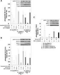
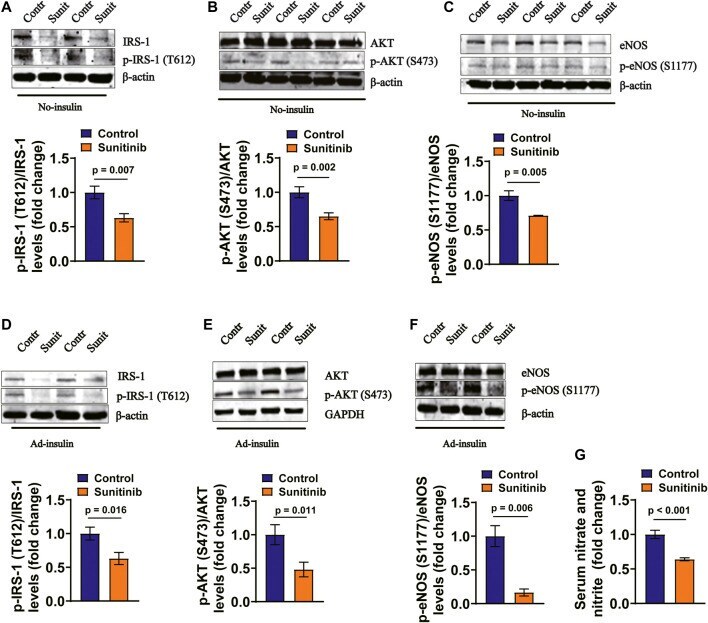
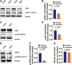
- FIGURE 4 Sunitinib inhibits insulin signaling pathway in mesenteric arteries in rats. (A) Representative western blots of total IRS-1 and phospho-Tyr612 IRS-1 in mesenteric artery tissues isolated on day 7 from control and sunitinib-treated rats, without insulin stimulation; bar graph represents phospho-Tyr 612 IRS-1 levels normalized to total IRS-1 levels (lower panel) ( p = 0.007 vs. control; n = 6 rats/group). (B) Representative western blots of total AKT and phospho-Ser473 AKT in mesenteric artery tissues isolated on day 7 from control and sunitinib-treated rats, without insulin stimulation; bar graph represents phospho-Ser473 AKT levels normalized to total AKT levels (lower panel) ( p = 0.002 vs. control; n = 7 rats/group). (C) Representative western blots of total eNOS and phospho-Ser1177 eNOS in mesenteric artery tissues isolated on day 7 from control and sunitinib-treated rats, without insulin stimulation; bar graph represents phospho-Ser1177 eNOS levels normalized to total eNOS levels (lower panel) ( p = 0.005 vs. control, n = 7 rats/group). (D) Representative western blot images of total IRS-1 and phospho-Tyr 612 IRS-1 in mesenteric artery tissues dissected on day 7 from insulin-injection control rats or insulin-injection rats treated with sunitinib; bar graph represents phospho-Tyr612 IRS-1levels normalized to total IRS-1 levels (lower panel) ( p = 0.016 vs. control; n = 7 per group). (E) Representative westerns of total AKT and phospho-Ser473 AKT in mesenteric art
- Submitted by
- Invitrogen Antibodies (provider)
- Main image
- Experimental details
- FIGURE 5 Sunitinib inhibits insulin signaling associated molecules in rat MAECs (A) Representative western blots of total IRS-1 and phospho-Tyr612 IRS-1 in control MAECs or in MAECs treated with 1 muM sunitinib for 30 min and bar graph represents phospho-Tyr612 IRS-1 levels normalized to total IRS-1 levels. (B) ( p = 0.015 vs. control; n = 6 batches of cells in each group). (C) Representative western blot images of total AKT and phospho-Ser473 AKT expression in control MAECs cells or in MAECs treated with 1 muM sunitinib for 30 min and bar graph represents phospho-Ser473 AKT levels normalized to total AKT levels. (D) ( p = 0.003 vs. control; n = 7 batches of cells in each group). (E) Representative western blot images of total eNOS and phospho-Ser1177 eNOS expression in control MAECs cell or in MAECs treated with 1 muM sunitinib for 30 min and bar graph represents phospho-Ser1177 eNOS levels normalized to total eNOS levels. (F) ( p = 0.002 vs. control, n = 7 batches of cells in each group). (G) The concentrations of nitrate and nitrite measured in the control MAECs cells or in MAECs treated with 1 muM sunitinib for 30 min, reflecting NO production under indicated experimental condition ( p < 0.001 vs. control; n = 6 per group).

- Submitted by
- Invitrogen Antibodies (provider)
- Main image
- Experimental details
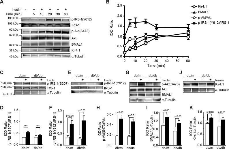
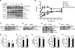
- Figure 6 Restoration of IRS-1 signaling corrects Kir4.1 levels. (A) rMC-1 cells were treated with 10 nM insulin, Western blot showing an increase in phosphorylation of IRS-1 Tyr612 , Akt Ser473 , BMAL-1, and Kir4.1. (B) Line chart showing quantification of the respective protein. The retinal explants from db/m and db/db mice were treated with insulin. (C) Western blot. (D) Bar chart showing an increase in phosphorylation of IRS-1 Ser307 . (E) Western blot showing IRS-1 Tyr612 phosphorylation after insulin treatment and (F) quantification of phospho-IRS-1 Tyr612 . (G) Western blot showing a concurrent increase in BMAL1 and phosphorylation of Akt Ser473 . (H) Bar chart showing a quantification of phospho-Akt Ser473 and (I) BMAL1. (J) Western blot showing Kir4.1 protein levels in insulin-treated explants. (K) Bar chart showing the quantification of Kir4.1 (n = 5).