Explore
 Validate
 Learn
Learn Western blot
 Immunocytochemistry
Antibody data
- Antibody Data
- Antigen structure
- References [2]
- Comments [0]
- Validations
- Immunocytochemistry [1]
- Immunohistochemistry [1]
Submit
Validation data
Reference
Comment
Report error
- Product number
- AMAb91251 - Provider product page

- Provider
- Atlas Antibodies
- Proper citation
- Atlas Antibodies Cat#AMAb91251, RRID:AB_2665863
- Product name
- Anti-LMNB1
- Antibody type
- Monoclonal
- Description
- Monoclonal Antibody against Human LMNB1, Clone ID: CL3929, Gene description: Lamin b1, Validated applications: WB, IHC, ICC, Uniprot ID: P20700, Storage: Store at +4°C for short term storage. Long time storage is recommended at -20°C.
- Reactivity
- Human
- Host
- Mouse
- Conjugate
- Unconjugated
- Isotype
- IgG
- Antibody clone number
- CL3929
- Vial size
- 100 µl
- Concentration
- 1.0 mg/ml
- Storage
- Store at +4°C for short term storage. Long time storage is recommended at -20°C.
- Handling
- The antibody solution should be gently mixed before use.
Submitted references An open source toolkit for repurposing Illumina sequencing systems as versatile fluidics and imaging platforms
Operation of a TCA cycle subnetwork in the mammalian nucleus.
Pandit K, Petrescu J, Cuevas M, Stephenson W, Smibert P, Phatnani H, Maniatis S
Scientific Reports 2022;12(1)
Scientific Reports 2022;12(1)
Operation of a TCA cycle subnetwork in the mammalian nucleus.
Kafkia E, Andres-Pons A, Ganter K, Seiler M, Smith TS, Andrejeva A, Jouhten P, Pereira F, Franco C, Kuroshchenkova A, Leone S, Sawarkar R, Boston R, Thaventhiran J, Zaugg JB, Lilley KS, Lancrin C, Beck M, Patil KR
Science advances 2022 Sep 2;8(35):eabq5206
Science advances 2022 Sep 2;8(35):eabq5206
No comments: Submit comment
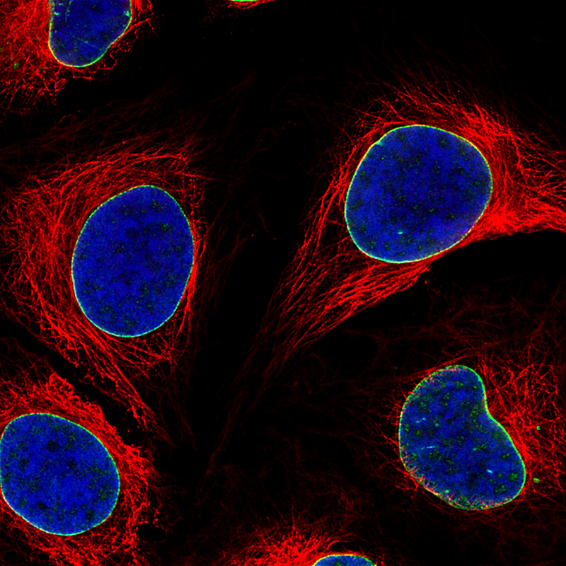
Supportive validation
- Submitted by
- Atlas Antibodies (provider)
- Main image

- Experimental details
- Immunofluorescence staining of MCF7 cells using the Anti-LMNB1 monoclonal antibody, showing specific staining of nuclear membrane in green. Microtubule- and nuclear probes are visualized in red and blue, respectively (where available).
- Sample type
- Human
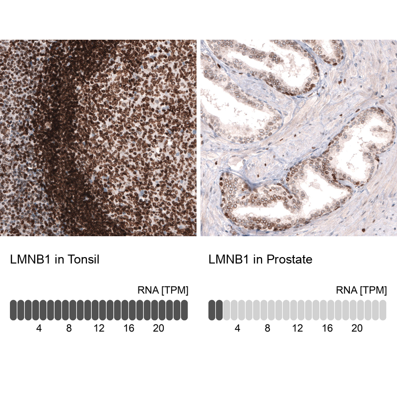
Supportive validation
- Submitted by
- Atlas Antibodies (provider)
- Enhanced method
- Orthogonal validation
- Main image
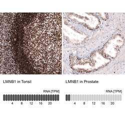
- Experimental details
- Immunohistochemistry analysis in human tonsil and prostate tissues using AMAb91251 antibody. Corresponding LMNB1 RNA-seq data are presented for the same tissues.
- Sample type
- Human
- Protocol
- Protocol