Explore
 Validate
 Learn
Learn Immunocytochemistry
Antibody data
- Antibody Data
- Antigen structure
- References [5]
- Comments [0]
- Validations
- Immunocytochemistry [3]
- Flow cytometry [2]
- Other assay [2]
Submit
Validation data
Reference
Comment
Report error
- Product number
- 11-1078-42 - Provider product page

- Provider
- Invitrogen Antibodies
- Product name
- CD107b (LAMP-2) Monoclonal Antibody (eBioH4B4 (H4B4)), FITC, eBioscience™
- Antibody type
- Monoclonal
- Antigen
- Other
- Description
- Description: The eBioH4B4 monoclonal antibody reacts with human CD107b, also known as lysosomal-associated membrane protein-2 (LAMP-2). CD107b is a highly glycosylated, type I transmembrane protein of approximately 105 kDa. It is expressed intracellularly in lysosomal/endosomal membranes in nearly all cells. It is also expressed on the surface of degranulating T cells (to a lesser extent than CD107a) and activated platelets as well as some cancer cells. In humans, mutations in CD107b results in a lysosomal glycogen storage disorder, known as Danon disease. Applications Reported: This eBioH4B4 (H4B4) antibody has been reported for use in intracellular staining followed by flow cytometric analysis. It has also been reported for use in surface staining in a flow cytometric based degranulation assay. Applications Tested: This eBioH4B4 (H4B4) antibody has been pre-titrated and tested by intracellular staining and flow cytometric analysis of Jurkat cells. This can be used at 5 µL (0.25 µg) per test. A test is defined as the amount (µg) of antibody that will stain a cell sample in a final volume of 100 µL. Cell number should be determined empirically but can range from 10^5 to 10^8 cells/test. Excitation: 488 nm; Emission: 520 nm; Laser: Blue Laser. Filtration: 0.2 µm post-manufacturing filtered.
- Reactivity
- Human
- Host
- Mouse
- Conjugate
- Green dye
- Isotype
- IgG
- Antibody clone number
- eBioH4B4 (H4B4)
- Vial size
- 100 Tests
- Concentration
- 5 μL/Test
- Storage
- 4°C, store in dark, DO NOT FREEZE!
Submitted references Improved Autophagic Flux in Escapers from Doxorubicin-Induced Senescence/Polyploidy of Breast Cancer Cells.
Lysosomal membrane permeabilization is involved in oxidative stress-induced apoptotic cell death in LAMP2-deficient iPSCs-derived cerebral cortical neurons.
Detection of T-cell degranulation: CD107a and b.
LAMP-1 and LAMP-2, but not LAMP-3, are reliable markers for activation-induced secretion of human mast cells.
Primary LAMP-2 deficiency causes X-linked vacuolar cardiomyopathy and myopathy (Danon disease).
Bojko A, Staniak K, Czarnecka-Herok J, Sunderland P, Dudkowska M, Śliwińska MA, Salmina K, Sikora E
International journal of molecular sciences 2020 Aug 24;21(17)
International journal of molecular sciences 2020 Aug 24;21(17)
Lysosomal membrane permeabilization is involved in oxidative stress-induced apoptotic cell death in LAMP2-deficient iPSCs-derived cerebral cortical neurons.
Law CY, Siu CW, Fan K, Lai WH, Au KW, Lau YM, Wong LY, Ho JCY, Lee YK, Tse HF, Ng KM
Biochemistry and biophysics reports 2016 Mar;5:335-345
Biochemistry and biophysics reports 2016 Mar;5:335-345
Detection of T-cell degranulation: CD107a and b.
Betts MR, Koup RA
Methods in cell biology 2004;75:497-512
Methods in cell biology 2004;75:497-512
LAMP-1 and LAMP-2, but not LAMP-3, are reliable markers for activation-induced secretion of human mast cells.
Grützkau A, Smorodchenko A, Lippert U, Kirchhof L, Artuc M, Henz BM
Cytometry. Part A : the journal of the International Society for Analytical Cytology 2004 Sep;61(1):62-8
Cytometry. Part A : the journal of the International Society for Analytical Cytology 2004 Sep;61(1):62-8
Primary LAMP-2 deficiency causes X-linked vacuolar cardiomyopathy and myopathy (Danon disease).
Nishino I, Fu J, Tanji K, Yamada T, Shimojo S, Koori T, Mora M, Riggs JE, Oh SJ, Koga Y, Sue CM, Yamamoto A, Murakami N, Shanske S, Byrne E, Bonilla E, Nonaka I, DiMauro S, Hirano M
Nature 2000 Aug 24;406(6798):906-10
Nature 2000 Aug 24;406(6798):906-10
No comments: Submit comment
Supportive validation
- Submitted by
- Invitrogen Antibodies (provider)
- Main image
- Experimental details
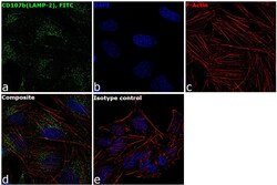
- Immunofluorescence analysis of CD107b (LAMP-2) was performed using 70% confluent log phase HeLa cells. The cells were fixed with 4% paraformaldehyde for 10 minutes, permeabilized with 0.1% Triton™ X-100 for 10 minutes, and blocked with 1% BSA for 1 hour at room temperature. The cells were labeled with CD107b(LAMP-2), FITC, Mouse Monoclonal antibody (Product # 11-1078-41) at 0.25 µg/mL in 0.1% BSA and incubated at 4 degree Celsius overnight (Panel a: green). Nuclei (Panel b: blue) were stained with SlowFade® Gold Antifade Mountant with DAPI (Product # S36938). F-actin (Panel c: red) was stained with Rhodamine Phalloidin (Product # R415, 1:300). Panel d represents the merged image showing cytoplasmic localization. Panel e represents the isotype control. The images were captured at 60X magnification.
- Submitted by
- Invitrogen Antibodies (provider)
- Main image
- Experimental details
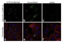
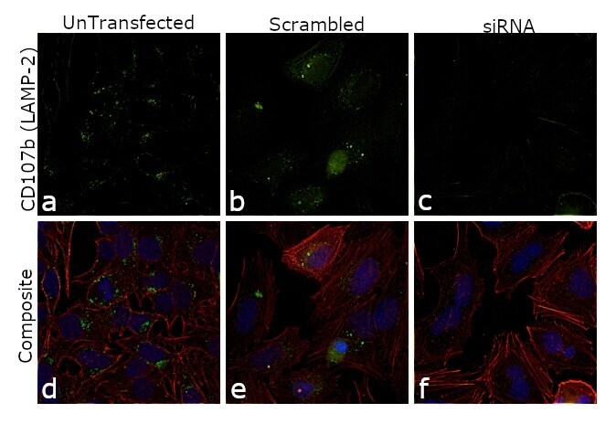
- Knockdown of CD107b (LAMP2) was achieved by transfecting HeLa cells with CD107b (LAMP2) specific siRNAs (Silencer® select Product # s533069, s8086 ). Immunofluorescence analysis was performed using untransfected HeLa cells (panels a, d), transfected with non-specific scrambled siRNA (panels b,e) and transfected with CD107b (LAMP2) specific siRNAs (panel c,f). Cells were fixed, permeabilized, and probed with CD107b (LAMP-2) Monoclonal Antibody (eBioH4B4 (H4B4)), FITC, eBioscience™ (Product # 11-1078-41, 1:250 dilution). Nuclei (blue) were stained using ProLong™ Diamond Antifade Mountant with DAPI (Product # P36962) and Rhodamine Phalloidin (Product # R415, 1:300) was used for cytoskeletal F-actin (red) staining. Reduction of specific cytoplasmic localization was observed upon siRNA mediated knockdown (panel c,f) confirming specificity of the antibody to CD107b (LAMP2). The images were captured at 60X magnification.
- Conjugate
- Green dye
- Submitted by
- Invitrogen Antibodies (provider)
- Main image

- Experimental details
- Knockdown of CD107b (LAMP2) was achieved by transfecting HeLa cells with CD107b (LAMP2) specific siRNAs (Silencer® select Product # s533069, s8086 ). Immunofluorescence analysis was performed using untransfected HeLa cells (panels a, d), transfected with non-specific scrambled siRNA (panels b,e) and transfected with CD107b (LAMP2) specific siRNAs (panel c,f). Cells were fixed, permeabilized, and probed with CD107b (LAMP-2) Monoclonal Antibody (eBioH4B4 (H4B4)), FITC, eBioscience™ (Product # 11-1078-41, 1:250 dilution). Nuclei (blue) were stained using ProLong™ Diamond Antifade Mountant with DAPI (Product # P36962) and Rhodamine Phalloidin (Product # R415, 1:300) was used for cytoskeletal F-actin (red) staining. Reduction of specific cytoplasmic localization was observed upon siRNA mediated knockdown (panel c,f) confirming specificity of the antibody to CD107b (LAMP2). The images were captured at 60X magnification.

Supportive validation
- Submitted by
- Invitrogen Antibodies (provider)
- Main image
- Experimental details
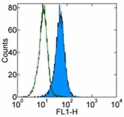
- Intracellular staining of the Jurkat cell line with Mouse IgG1 K Isotype Control FITC (Product # 11-4714-42) (open histogram) or Anti-Human CD107b (LAMP-2) FITC (filled histogram). Total cells were used for analysis.
- Conjugate
- Green dye

- Submitted by
- Invitrogen Antibodies (provider)
- Main image

- Experimental details
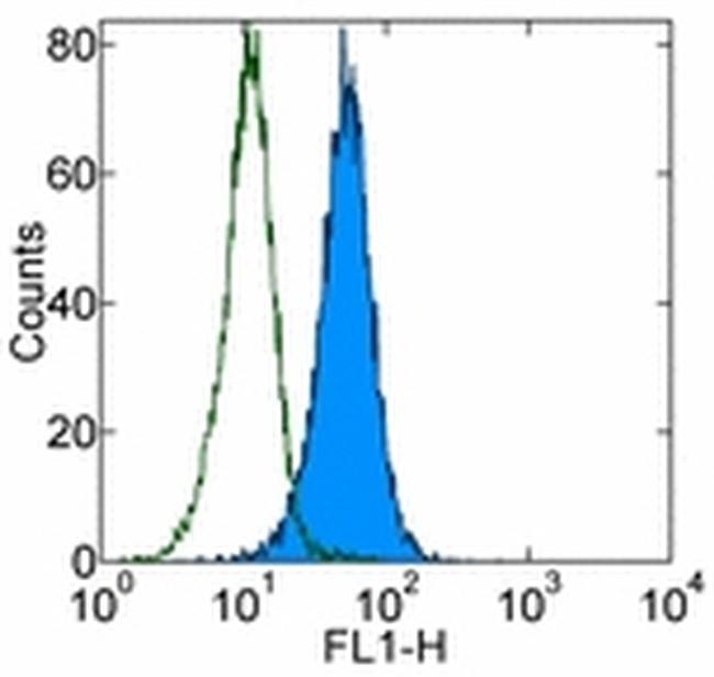
- Intracellular staining of the Jurkat cell line with Mouse IgG1 K Isotype Control FITC (Product # 11-4714-42) (open histogram) or Anti-Human CD107b (LAMP-2) FITC (filled histogram). Total cells were used for analysis.
Supportive validation
- Submitted by
- Invitrogen Antibodies (provider)
- Main image
- Experimental details
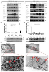
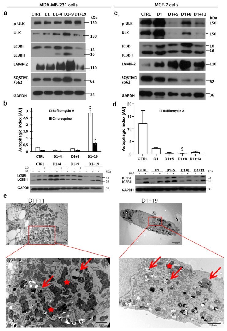
- Figure 5 Autophagic flux resumption in descendants of polyploid/senescent MDA-MB-231 and MCF-7 cells. Cells were treated with 100 nM doxorubicin for one day, then cultured in a fresh medium and analyzed on subsequent days. ( a , c ) Representative western blots showing autophagy protein levels of p-ULK1 (S757), ULK1, LC3B, LAMP-2 and SQSTM1/p62 in MDA-MB-231 cells ( a ) and MCF-7 cells ( c ). ( b , d ) Quantitative analysis of autophagic index based on LC3B protein levels in untreated and bafilomycin A- or chloroquine-treated MDA-MB-231 cells ( b ) and MCF-7 cells ( d ) with representative western blots showing LC3B protein levels. Bars: mean value, error bars: SEM, n = 4. Statistical significance (in relation to control): $ p < 0.051, * p < 0.05, ** p < 0.01. ( e ) Transient accumulation of autophagic vesicles. TEM images show typical MDA-MB-231cells on the subsequent days following treatment (upper panel) and their magnified parts with autophagic vesicles (red arrows) and lipofuscin particles (red asterisks).
- Conjugate
- Green dye
- Submitted by
- Invitrogen Antibodies (provider)
- Main image

- Experimental details
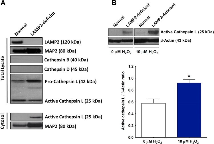
- Fig. 4 LAMP2-deficient neurons showed increase levels of cytosolic cathepsin L. (A) Western blot analysis revealed the elevated level of active cathepsin L in the LAMP2-deficient iPSCs-derived cortical neurons under non-stressed condition. (B) Western blot analysis showed that oxidative stress increased the abundance of active cathepsin L in the cytosols. N =3; *: P
- Conjugate
- Green dye