 Explore
 Validate
 Learn
Learn Western blot
Antibody data
- Antibody Data
- Antigen structure
- References [0]
- Comments [0]
- Validations
- Western blot [2]
- Immunocytochemistry [1]
- Flow cytometry [1]
Submit
Validation data
Reference
Comment
Report error
- Product number
- MA1-165 - Provider product page

- Provider
- Invitrogen Antibodies
- Product name
- LAMP2 Monoclonal Antibody (GL2A7)
- Antibody type
- Monoclonal
- Antigen
- Purifed from natural sources
- Description
- MA1-165 detects LAMP2 in mouse samples. MA1-165 has been successfully used in Western blot, immunofluorescence and flow cytometry applications. The MA1-165 immunogen is a purified preparation of mouse liver lysosomal membranes.
- Reactivity
- Human, Mouse, Rat
- Host
- Rat
- Isotype
- IgG
- Antibody clone number
- GL2A7
- Vial size
- 100 µL
- Concentration
- 1 mg/mL
- Storage
- -20° C, Avoid Freeze/Thaw Cycles
No comments: Submit comment
Supportive validation
- Submitted by
- Invitrogen Antibodies (provider)
- Main image
- Experimental details
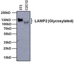
- Western blot analysis of LAMP2 was performed by loading 20 µg of the indicated whole cell lysates and 5 µL of PageRuler Plus Prestained Protein Ladder (Product # 26619) per well onto a Novex 4-20% Tris-Glycine polyacrylamide gel (Product # WT4202BOX ). Proteins were transferred to a nitrocellulose membrane using the G2 Blotter (Product # 62288), and blocked with 5% Milk in TBST for 1 hour at room temperature. LAMP2 was detected at ~100-130 kDa using a LAMP2 monoclonal antibody (Product # MA1-165) at a dilution of 1:1000 in 5% Milk in TBST overnight at 4C on a rocking platform, followed by a Goat anti-Rat IgG Secondary Antibody, HRP conjugate (Product # 31470) at a dilution of 1:10,000 for at least 30 minutes at room temperature. Chemiluminescent detection was performed using SuperSignal Pico substrate (Product # 34078) and the myECL Imager (Product # 62236).

- Submitted by
- Invitrogen Antibodies (provider)
- Main image
- Experimental details
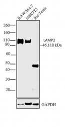
- Western blot analysis was performed on membrane enriched extracts (30 µg lysate) of RAW 264.7 (Lane 1), NIH/3T3 (Lane 2) and tissue extract (30 µg lysate) of Rat Testis (Lane 3). The blot was probed with Anti-LAMP2 Monoclonal Antibody (Product # MA5-17862, 1:1000 dilution) and detected by chemiluminescence using Sheep anti-Rat IgG Secondary Antibody, HRP conjugate (Product # PA1-28642, 0.2 µg/mL, 1:5000 dilution). A 46 kDa band corresponding to LAMP2 was observed in Rat Testis. Apart from the desired band, a 110 kDa band was observed across cell lines which is the glycosylated form of LAMP2. Known quantity of protein samples were electrophoresed using Novex® NuPAGE® 4-12 % Bis-Tris gel (Product # NP0322BOX), XCell SureLock™ Electrophoresis System (Product # EI0002) and Novex® Sharp Pre-Stained Protein Standard (Product # LC5800). Resolved proteins were then transferred onto a nitrocellulose membrane with iBlot® 2 Dry Blotting System (Product # IB21001). The membrane was probed with the relevant primary and secondary Antibody following blocking with 5% skimmed milk. Chemiluminescent detection was performed using Pierce™ ECL Western Blotting Substrate (Product # 32106).

Supportive validation
- Submitted by
- Invitrogen Antibodies (provider)
- Main image

- Experimental details
- Immunofluorescent analysis of LAMP2 (green) in 3T3 cells. The cells were fixed with 4% paraformaldehyde for 15 minutes, permeabilized with 0.1% Triton X-100 in PBS for 15 minutes, and blocked with 3% BSA in PBS (Product # 37525) for 30 minutes at room temperature. Cells were stained with a LAMP2 monoclonal antibody (Product # MA1-165) at a dilution of 10 µg/mL in staining buffer for 1 hour at room temperature, and then incubated with a Goat anti-Rat IgG Secondary Antibody, DyLight 488 conjugate (Product # SA5-10018) at a dilution of 1:1000 for 1 hour at room temperature (green). Nuclei (blue) were counterstained with Hoechst 33342 dye (Product # 62249). Images were taken on a Thermo Scientific ToxInsight Instrument at 20X magnification.

Supportive validation
- Submitted by
- Invitrogen Antibodies (provider)
- Main image

- Experimental details
- Flow cytometry analysis of LAMP2 was done on 3T3 cells. Cells were fixed, permeabilized and stained with a LAMP2 mouse monoclonal antibody (Product # MA1-165, orange histogram) or an isotype control (Product # 02-9602, black histogram) at a dilution of 2.5 µg/mL. After incubation for 1 hour on ice, the cells were labeled with Goat anti-Rat IgG Secondary Antibody, DyLight 488 conjugate (Product # SA5-10018) at a dilution of 1:50 for 1 hour on ice. A representative 10,000 cells were acquired and analyzed for each sample.
