Explore
 Validate
 Learn
Learn Western blot
Antibody data
- Antibody Data
- Antigen structure
- References [6]
- Comments [0]
- Validations
- Western blot [1]
- Immunocytochemistry [2]
- Flow cytometry [1]
Submit
Validation data
Reference
Comment
Report error
- Product number
- MA1-205 - Provider product page

- Provider
- Invitrogen Antibodies
- Product name
- LAMP2 Monoclonal Antibody (H4B4)
- Antibody type
- Monoclonal
- Antigen
- Other
- Description
- MA1-205 Lamp2 antibody detects Lamp2 by western blot, immunofluorescence and flow cytometry. MA1-205 Lamp2 antibody detects Lamp2 by western blot at ~115kda which is larger than the protein itself due to heavy glycosylation.
- Reactivity
- Human, Mouse
- Host
- Mouse
- Isotype
- IgG
- Antibody clone number
- H4B4
- Vial size
- 100 µg
- Concentration
- 1 mg/mL
- Storage
- -20°C
Submitted references Exosome-mediated mRNA delivery in vivo is safe and can be used to induce SARS-CoV-2 immunity.
Efficient Protein Transfection by Swarms of Chemically Powered Plasmonic Virus-Sized Nanorobots.
KIF5A-dependent axonal transport deficiency disrupts autophagic flux in trimethyltin chloride-induced neurotoxicity.
Inhibiting autophagy targets human leukemic stem cells and hypoxic AML blasts by disrupting mitochondrial homeostasis.
The D614G Mutation Enhances the Lysosomal Trafficking of SARS-CoV-2 Spike.
The Tumor Antigen NY-ESO-1 Mediates Direct Recognition of Melanoma Cells by CD4+ T Cells after Intercellular Antigen Transfer.
Tsai SJ, Atai NA, Cacciottolo M, Nice J, Salehi A, Guo C, Sedgwick A, Kanagavelu S, Gould SJ
The Journal of biological chemistry 2021 Nov;297(5):101266
The Journal of biological chemistry 2021 Nov;297(5):101266
Efficient Protein Transfection by Swarms of Chemically Powered Plasmonic Virus-Sized Nanorobots.
Ressnerova A, Novotny F, Michalkova H, Pumera M, Adam V, Heger Z
ACS nano 2021 Aug 24;15(8):12899-12910
ACS nano 2021 Aug 24;15(8):12899-12910
KIF5A-dependent axonal transport deficiency disrupts autophagic flux in trimethyltin chloride-induced neurotoxicity.
Liu M, Pi H, Xi Y, Wang L, Tian L, Chen M, Xie J, Deng P, Zhang T, Zhou C, Liang Y, Zhang L, He M, Lu Y, Chen C, Yu Z, Zhou Z
Autophagy 2021 Apr;17(4):903-924
Autophagy 2021 Apr;17(4):903-924
Inhibiting autophagy targets human leukemic stem cells and hypoxic AML blasts by disrupting mitochondrial homeostasis.
Dykstra KM, Fay HRS, Massey AC, Yang N, Johnson M, Portwood S, Guzman ML, Wang ES
Blood advances 2021 Apr 27;5(8):2087-2100
Blood advances 2021 Apr 27;5(8):2087-2100
The D614G Mutation Enhances the Lysosomal Trafficking of SARS-CoV-2 Spike.
Guo C, Tsai SJ, Ai Y, Li M, Pekosz A, Cox A, Atai N, Gould SJ
bioRxiv : the preprint server for biology 2020 Dec 9;
bioRxiv : the preprint server for biology 2020 Dec 9;
The Tumor Antigen NY-ESO-1 Mediates Direct Recognition of Melanoma Cells by CD4+ T Cells after Intercellular Antigen Transfer.
Fonteneau JF, Brilot F, Münz C, Gannagé M
Journal of immunology (Baltimore, Md. : 1950) 2016 Jan 1;196(1):64-71
Journal of immunology (Baltimore, Md. : 1950) 2016 Jan 1;196(1):64-71
No comments: Submit comment
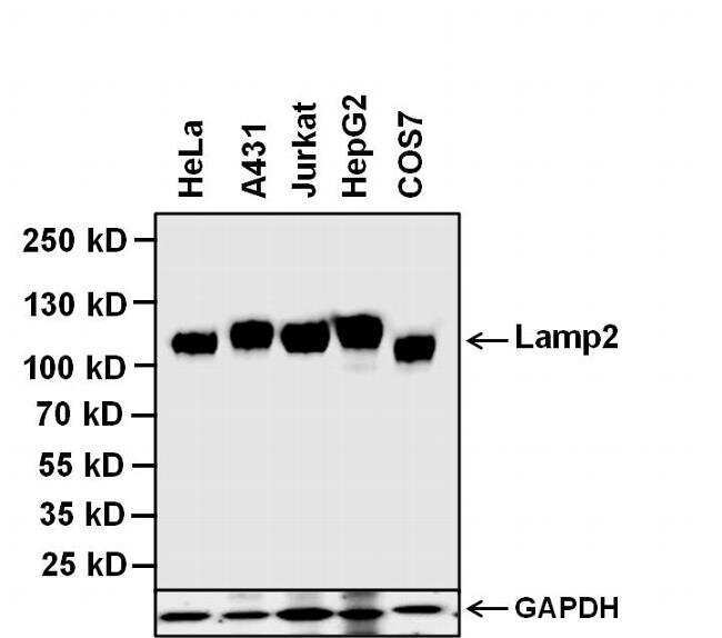
Supportive validation
- Submitted by
- Invitrogen Antibodies (provider)
- Main image

- Experimental details
- Western blot analysis of LAMP-2 was performed by loading 20 µg of the indicated whole cell lysates and 5 µL of PageRuler Plus Prestained Protein Ladder (Product # 26619) per well onto a 4-20% Tris-Glycine polyacrylamide gel (Product # WT4202BX10). Proteins were transferred to a nitrocellulose membrane using the G2 Blotter (Product # 62288), and blocked with 5% Milk in TBST for 1 hour at room temperature. LAMP-2 was detected at ~110 kDa using a LAMP-2 mouse monoclonal antibody (Product # MA1-205) at a dilution of 1 µg/mL in blocking buffer overnight at 4 C on a rocking platform, followed by a Goat anti-Mouse IgG (H+L) Superclonal™ Secondary Antibody, HRP conjugate (Product # A28177) at a dilution of 1:1000 for at least 30 minutes at room temperature. Chemiluminescent detection was performed using SuperSignal West Pico (Product # 34078). GAPDH was detected GAPDH rabbit polyclonal antibody, HRP conjugate (Product # PA1-987-HRP), at a dilution of 1:1000 for at least 1 hour at room temperature.
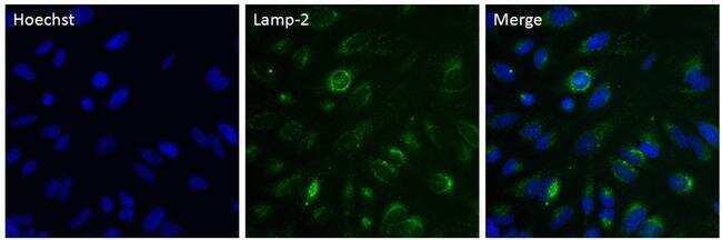
Supportive validation
- Submitted by
- Invitrogen Antibodies (provider)
- Main image

- Experimental details
- Immunofluorescent analysis of LAMP-2 in HeLa cells. The cells were fixed with 4% paraformaldehyde in PBS for 15 minutes, permeabilized with 0.1% Triton X-100 for 15 minutes, and blocked with 3% BSA in PBS for 30 minutes at room temperature. Cells were stained with a LAMP-2 mouse monoclonal antibody (Product # MA1-205) at a dilution of 5 µg/mL in blocking buffer for 1 hour at room temperature, and then incubated with Goat anti-Mouse IgG (H+L) Superclonal™ Secondary Antibody, Alexa Fluor® 488 conjugate (Product # A28175) at a dilution of 1:500 for at least 30 minutes at room temperature in the dark (green). Nuclei (blue) were stained with Hoechst 33342 (Product # 62249). Images were taken on a Thermo Scientific ToxInsight Instrument at 20X magnification.
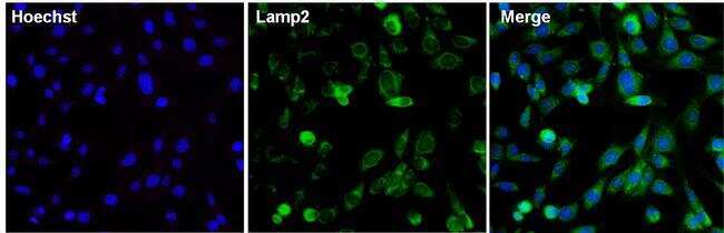
- Submitted by
- Invitrogen Antibodies (provider)
- Main image

- Experimental details
- Immunofluorescent analysis of LAMP-2 in 3T3 cells. The cells were fixed with 4% paraformaldehyde in PBS for 15 minutes, permeabilized with 0.1% Triton X-100 for 15 minutes, and blocked with 3% BSA in PBS for 30 minutes at room temperature. Cells were stained with a LAMP-2 mouse monoclonal antibody (Product # MA1-205) at a dilution of 5 µg/mL in blocking buffer for 1 hour at room temperature, and then incubated with Goat anti-Mouse IgG (H+L) Superclonal™ Secondary Antibody, Alexa Fluor® 488 conjugate (Product # A28175) at a dilution of 1:500 for at least 30 minutes at room temperature in the dark (green). Nuclei (blue) were stained with Hoechst 33342 (Product # 62249). Images were taken on a Thermo Scientific ToxInsight Instrument at 20X magnification.
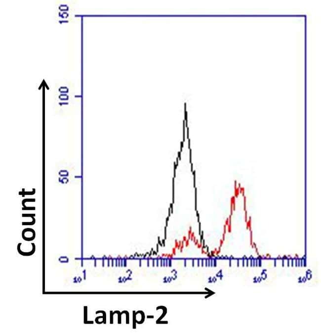
Supportive validation
- Submitted by
- Invitrogen Antibodies (provider)
- Main image

- Experimental details
- Flow cytometry analysis of LAMP-2 was done on HeLa cells. Cells were fixed, permeabilized and stained with a LAMP-2 mouse monoclonal antibody (Product # MA1-205, red histogram) at a dilution of 10 µg/mL. After incubation of the primary antibody on ice for 1 hour, the cells were stained with a Goat anti-Mouse IgG (H+L) Secondary Antibody, DyLight 680 conjugate (Product # 35519) at a dilution of 1:50 for at least 30 minutes on ice. A representative 10,000 cells were acquired for each sample. The black histogram represents unstained control cells and the blue histogram represents no-primary-antibody control.