 Explore
 Validate
 Learn
Learn Immunocytochemistry
 Immunohistochemistry
Antibody data
- Antibody Data
- Antigen structure
- References [0]
- Comments [0]
- Validations
- Immunohistochemistry [1]
- Flow cytometry [3]
Submit
Validation data
Reference
Comment
Report error
- Product number
- NBP2-44306 - Provider product page

- Provider
- Novus Biologicals
- Product name
- Mouse Monoclonal HGFR/c-MET Antibody
- Antibody type
- Monoclonal
- Description
- Protein G purified.
- Reactivity
- Human
- Host
- Mouse
- Isotype
- IgG
- Vial size
- 0.1 mg
- Concentration
- 1.0 mg/ml
- Storage
- Store at 4C short term. Aliquot and store at -20C long term. Avoid freeze-thaw cycles.
No comments: Submit comment
Supportive validation
- Submitted by
- Novus Biologicals (provider)
- Main image
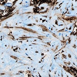
- Experimental details
- Immunohistochemistry-Paraffin: HGFR/c-MET Antibody (1G7NB) [NBP2-44306] - Analysis of HGF R/c-Met (1G7NB) in human liver cancer.

Supportive validation
- Submitted by
- Novus Biologicals (provider)
- Main image
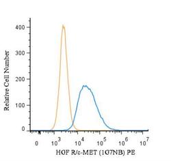
- Experimental details
- Flow (Intracellular): HGF R/c-MET Antibody (1G7NB) [NBP2-44306] - An intracellular stain was performed on HeLa cells with NBP2-44306PE (blue) and a matched isotype control (orange). Cells were fixed with 4% PFA and then permeabilized with 0.1% saponin. Cells were incubated in an antibody dilution of 5 ug/mL for 30 minutes at room temperature. Both antibodies were conjugated to phycoerythrin.

- Submitted by
- Novus Biologicals (provider)
- Main image

- Experimental details
- Flow (Intracellular): HGF R/c-MET Antibody (1G7NB) [NBP2-44306] - An intracellular stain was performed on HeLa cells with HGF R/c-MET [1G7NB] Antibody NBP2-44306AF647 (blue) and a matched isotype control (orange). Cells were fixed with 4% PFA and then permeabilized with 0.1% saponin. Cells were incubated in an antibody dilution of 2.5 ug/mL for 30 minutes at room temperature. Both antibodies were conjugated to Alexa Fluor 647.

- Submitted by
- Novus Biologicals (provider)
- Main image

- Experimental details
- Flow Cytometry: HGFR/c-MET Antibody (1G7NB) [NBP2-44306] - c-MET antibody was tested in HeLa cells (1 x 10^6 cells/ml). After fixation and permeabilization, cells were stained using the anti-c-MET antibody (clone 1G7NB) at a 1:1000 dilution. Signal was detected using a Goat anti-Mouse Dylight 488 secondary (blue peak). Shown with secondary control (orange peak). Data was acquired on BD FACSCalibur. Image using the Azide Free format of this antibody.
