Explore
 Validate
 Learn
Learn Western blot
Antibody data
- Antibody Data
- Antigen structure
- References [7]
- Comments [0]
- Validations
- Western blot [2]
- Immunocytochemistry [1]
- Immunohistochemistry [2]
- Flow cytometry [1]
Submit
Validation data
Reference
Comment
Report error
- Product number
- AF2480 - Provider product page

- Provider
- R&D Systems
- Product name
- Human/Mouse Phospho-HGFR/c-MET (Y1234/Y1235) Antibody
- Antibody type
- Polyclonal
- Description
- Antigen Affinity-purified. Detects human and mouse HGF R/c-MET when phosphorylated at Y1234/Y1235 in Western blots.
- Reactivity
- Human, Mouse
- Host
- Rabbit
- Conjugate
- Unconjugated
- Isotype
- IgG
- Vial size
- 50 ug
- Concentration
- LYOPH
- Storage
- Use a manual defrost freezer and avoid repeated freeze-thaw cycles. 12 months from date of receipt, -20 to -70 °C as supplied. 1 month, 2 to 8 °C under sterile conditions after reconstitution. 6 months, -20 to -70 °C under sterile conditions after reconstitution.
Submitted references Reviving oncogenic addiction to MET bypassed by BRAF (G469A) mutation.
Pulmonary pericytes regulate lung morphogenesis.
Dual MET/EGFR therapy leads to complete response and resistance prevention in a MET-amplified gastroesophageal xenopatient cohort.
Targeting MET kinase with the small-molecule inhibitor amuvatinib induces cytotoxicity in primary myeloma cells and cell lines.
Protein signatures for classification and prognosis of gastric cancer a signaling pathway-based approach.
Alternative proteolytic processing of hepatocyte growth factor during wound repair.
Combined inhibition of MET and EGFR suppresses proliferation of malignant mesothelioma cells.
Virzì AR, Gentile A, Benvenuti S, Comoglio PM
Proceedings of the National Academy of Sciences of the United States of America 2018 Oct 2;115(40):10058-10063
Proceedings of the National Academy of Sciences of the United States of America 2018 Oct 2;115(40):10058-10063
Pulmonary pericytes regulate lung morphogenesis.
Kato K, Diéguez-Hurtado R, Park DY, Hong SP, Kato-Azuma S, Adams S, Stehling M, Trappmann B, Wrana JL, Koh GY, Adams RH
Nature communications 2018 Jun 22;9(1):2448
Nature communications 2018 Jun 22;9(1):2448
Dual MET/EGFR therapy leads to complete response and resistance prevention in a MET-amplified gastroesophageal xenopatient cohort.
Apicella M, Migliore C, Capelôa T, Menegon S, Cargnelutti M, Degiuli M, Sapino A, Sottile A, Sarotto I, Casorzo L, Cassoni P, De Simone M, Comoglio PM, Marsoni S, Corso S, Giordano S
Oncogene 2017 Mar 2;36(9):1200-1210
Oncogene 2017 Mar 2;36(9):1200-1210
Targeting MET kinase with the small-molecule inhibitor amuvatinib induces cytotoxicity in primary myeloma cells and cell lines.
Phillip CJ, Zaman S, Shentu S, Balakrishnan K, Zhang J, Baladandayuthapani V, Taverna P, Redkar S, Wang M, Stellrecht CM, Gandhi V
Journal of hematology & oncology 2013 Dec 10;6:92
Journal of hematology & oncology 2013 Dec 10;6:92
Protein signatures for classification and prognosis of gastric cancer a signaling pathway-based approach.
Wang D, Ye F, Sun Y, Li W, Liu H, Jiang J, Zhang Y, Liu C, Tong W, Gao L, Sun Y, Zhang W, Seetoe T, Lee P, Suo J, Zhang DY
The American journal of pathology 2011 Oct;179(4):1657-66
The American journal of pathology 2011 Oct;179(4):1657-66
Alternative proteolytic processing of hepatocyte growth factor during wound repair.
Buchstein N, Hoffmann D, Smola H, Lang S, Paulsson M, Niemann C, Krieg T, Eming SA
The American journal of pathology 2009 Jun;174(6):2116-28
The American journal of pathology 2009 Jun;174(6):2116-28
Combined inhibition of MET and EGFR suppresses proliferation of malignant mesothelioma cells.
Kawaguchi K, Murakami H, Taniguchi T, Fujii M, Kawata S, Fukui T, Kondo Y, Osada H, Usami N, Yokoi K, Ueda Y, Yatabe Y, Ito M, Horio Y, Hida T, Sekido Y
Carcinogenesis 2009 Jul;30(7):1097-105
Carcinogenesis 2009 Jul;30(7):1097-105
No comments: Submit comment
Supportive validation
- Submitted by
- R&D Systems (provider)
- Main image
- Experimental details
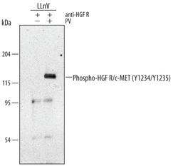
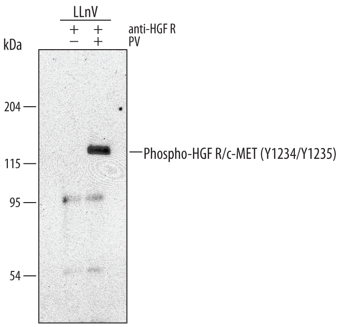
- Detection of Human Phospho-HGF R/c-MET (Y1234/Y1235) by Western Blot. Western blot shows Goat Anti-Human HGF R/c-MET Antigen Affinity-purified Polyclonal Antibody (Catalog # AF276) immunoprecipitate of MDA-MB-468 human breast cancer cell line untreated (-) or treated (+) with 100 μM pervanadate (PV) for 10 minutes. PVDF membrane was probed with 0.5 µg/mL of Rabbit Anti-Human/Mouse Phospho-HGF R/c-MET (Y1234/Y1235) Antigen Affinity-purified Polyclonal Antibody (Catalog # AF2480), followed by HRP-conjugated Anti-Rabbit IgG Secondary Antibody (Catalog # HAF008). A specific band was detected for Phospho-HGF R/c-MET (Y1234/Y1235) at approximately 145 kDa (as indicated). This experiment was conducted under reducing conditions and using Immunoblot Buffer Group 1.
- Submitted by
- R&D Systems (provider)
- Main image
- Experimental details
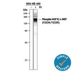
- Detection of Human Phospho-HGF R/c-MET (Y1234/Y1235) by Simple WesternTM. Simple Western lane view shows lysates of MDA-MB-468 human breast cancer cell line untreated (-) or treated (+) with 100 µM Pervanadate (PV) for 10 minutes, loaded at 0.2 mg/mL. A specific band was detected for HGF R/c-MET at approximately 156 kDa (as indicated) using 5 µg/mL of Rabbit Anti-Human/Mouse Phospho-HGF R/c-MET (Y1234/Y1235) Antigen Affinity-purified Polyclonal Antibody (Catalog # AF2480). This experiment was conducted under reducing conditions and using the 12-230 kDa separation system.

Supportive validation
- Submitted by
- R&D Systems (provider)
- Main image

- Experimental details
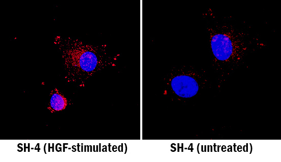
- HGF R/c-MET in SH-4 Human Cell Line. HGF R/c-MET was detected in immersion fixed SH-4 human melanoma cell line stimulated with HGF (left panel; positive staining) and non-stimulated (right panel; negative staining) using Rabbit Anti-Human/Mouse Phospho-HGF R/c-MET (Y1234/Y1235) Antigen Affinity-purified Polyclonal Antibody (Catalog # AF2480) at 15 µg/mL for 3 hours at room temperature. Cells were stained using the NorthernLights™ 557-conjugated Anti-Rabbit IgG Secondary Antibody (red; Catalog # NL004) and counterstained with DAPI (blue). Specific staining was localized to cytoplasm. View our protocol for Fluorescent ICC Staining of Cells on Coverslips.
Supportive validation
- Submitted by
- R&D Systems (provider)
- Main image

- Experimental details
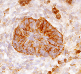
- HGF R/c-MET in Mouse Embryo. HGF R/c-MET was detected in immersion fixed frozen sections of mouse embryo (13 d.p.c.) using Rabbit Anti-Human/Mouse Phospho-HGF R/c-MET (Y1234/Y1235) Antigen Affinity-purified Polyclonal Antibody (Catalog # AF2480) at 15 µg/mL overnight at 4 °C. Tissue was stained using the Anti-Rabbit HRP-DAB Cell & Tissue Staining Kit (brown; Catalog # CTS005) and counterstained with hematoxylin (blue). View our protocol for Chromogenic IHC Staining of Frozen Tissue Sections.
- Submitted by
- R&D Systems (provider)
- Main image
- Experimental details
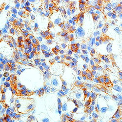
- HGF R/c-MET in Human Renal Cell Carcinoma Tissue. HGF R/c-MET was detected in immersion fixed paraffin-embedded sections of human renal cell carcinoma tissue using Rabbit Anti-Human/Mouse Phospho-HGF R/c-MET (Y1234/Y1235) Antigen Affinity-purified Polyclonal Antibody (Catalog # AF2480) at 15 µg/mL for 1 hour at room temperature followed by incubation with the Anti-Rabbit IgG VisUCyte™ HRP Polymer Antibody (Catalog # VC003). Before incubation with the primary antibody, tissue was subjected to heat-induced epitope retrieval using Antigen Retrieval Reagent-Basic (Catalog # CTS013). Tissue was stained using DAB (brown) and counterstained with hematoxylin (blue). Specific staining was localized to cytoplasm. View our protocol for IHC Staining with VisUCyte HRP Polymer Detection Reagents.

Supportive validation
- Submitted by
- R&D Systems (provider)
- Main image
- Experimental details
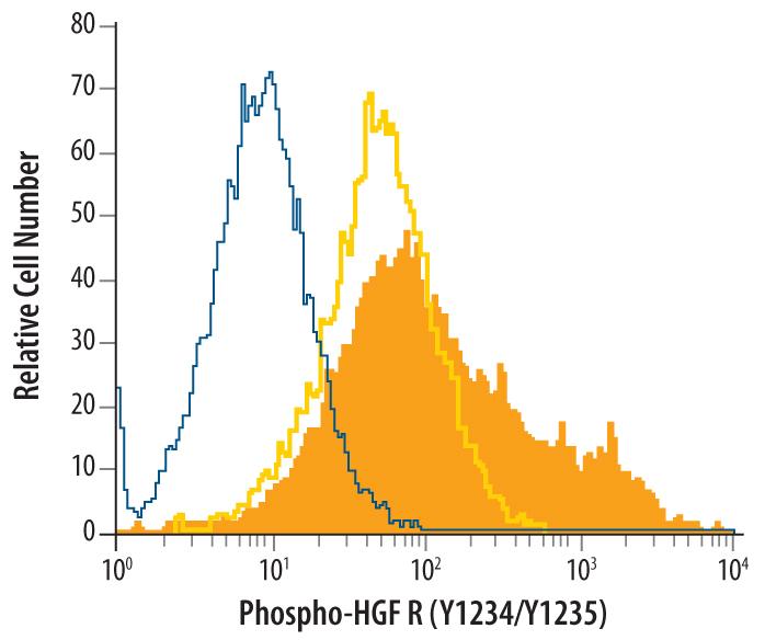
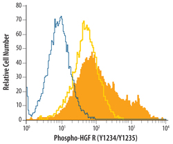
- Detection of HGF R/c-MET in pervanadate-treated MCF-7 Human Cell Line by Flow Cytometry. MCF-7 human breast cancer cell line was unstimulated (light orange open histogram) or treated with 100 μM pervanadate for 10 minutes (dark orange filled histogram), then stained with Rabbit Anti-Human/Mouse Phospho-HGF R/c-MET (Y1234/Y1235) Antigen Affinity-purified Polyclonal Antibody (Catalog # AF2480), or control antibody (Catalog # AB-105-C, blue open histogram), followed by Phycoerythrin-conjugated Anti-Rabbit IgG Secondary Antibody (Catalog # F0110). To facilitate intracellular staining, cells were fixed with paraformaldehyde and permeabilized with methanol.