Explore
 Validate
 Learn
Learn Western blot
 Immunocytochemistry
 Immunoprecipitation
Antibody data
- Antibody Data
- Antigen structure
- References [13]
- Comments [0]
- Validations
- Immunocytochemistry [5]
- Immunohistochemistry [2]
- Other assay [5]
Submit
Validation data
Reference
Comment
Report error
- Product number
- PA1-887 - Provider product page

- Provider
- Invitrogen Antibodies
- Product name
- MECP2 Polyclonal Antibody
- Antibody type
- Polyclonal
- Antigen
- Synthetic peptide
- Description
- PA1-887 detects methyl CpG binding protein 2 (MeCP2) from human, mouse and rat tissues and cells. PA1-887 has been successfully used in Western blot, IHC-P, immunofluorescence and immunoprecipitation procedures. By Western blot, this antibody detects an ~56 kDa protein representing MeCP2 from AtT20 cell extract. PA1-887 immunizing peptide corresponds to amino acid residues 1-15 from mouse MeCP2. This sequence is completely conserved in human MeCP2. PA1-887 immunizing peptide (Cat. # PEP-120) is available for use in neutralization and control experiments.
- Reactivity
- Human, Mouse, Rat
- Host
- Rabbit
- Isotype
- IgG
- Vial size
- 50 μg
- Concentration
- 1 mg/mL
- Storage
- -20°C, Avoid Freeze/Thaw Cycles
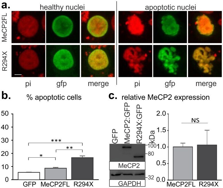
Submitted references Apoptotic Activity of MeCP2 Is Enhanced by C-Terminal Truncating Mutations.
Rett Syndrome Mutant Neural Cells Lacks MeCP2 Immunoreactive Bands.
MECP2 impairs neuronal structure by regulating KIBRA.
The Effects of Maternal Separation on Adult Methamphetamine Self-Administration, Extinction, Reinstatement, and MeCP2 Immunoreactivity in the Nucleus Accumbens.
Regulation of intestinal stem cell proliferation by human methyl-CpG-binding protein-2 in Drosophila.
The first missense mutation causing Rett syndrome specifically affecting the MeCP2_e1 isoform.
Genetic modifiers of MeCP2 function in Drosophila.
RNAi-induced down-regulation of Mecp2 expression in the rat brain.
Multiple pathways regulate MeCP2 expression in normal brain development and exhibit defects in autism-spectrum disorders.
A WW domain binding region in methyl-CpG-binding protein MeCP2: impact on Rett syndrome.
Quantitative localization of heterogeneous methyl-CpG-binding protein 2 (MeCP2) expression phenotypes in normal and Rett syndrome brain by laser scanning cytometry.
Quantitative localization of heterogeneous methyl-CpG-binding protein 2 (MeCP2) expression phenotypes in normal and Rett syndrome brain by laser scanning cytometry.
Methylated DNA and MeCP2 recruit histone deacetylase to repress transcription.
Williams AA, Mehler VJ, Mueller C, Vonhoff F, White R, Duch C
PloS one 2016;11(7):e0159632
PloS one 2016;11(7):e0159632
Rett Syndrome Mutant Neural Cells Lacks MeCP2 Immunoreactive Bands.
Bueno C, Tabares-Seisdedos R, Moraleda JM, Martinez S
PloS one 2016;11(4):e0153262
PloS one 2016;11(4):e0153262
MECP2 impairs neuronal structure by regulating KIBRA.
Williams AA, White R, Siniard A, Corneveaux J, Huentelman M, Duch C
Neurobiology of disease 2016 Jul;91:284-91
Neurobiology of disease 2016 Jul;91:284-91
The Effects of Maternal Separation on Adult Methamphetamine Self-Administration, Extinction, Reinstatement, and MeCP2 Immunoreactivity in the Nucleus Accumbens.
Lewis CR, Staudinger K, Scheck L, Olive MF
Frontiers in psychiatry 2013;4:55
Frontiers in psychiatry 2013;4:55
Regulation of intestinal stem cell proliferation by human methyl-CpG-binding protein-2 in Drosophila.
Lee SH, Kim IJ, Kim JG, Park JS, Kim YS, Yamaguchi M, Kim CM, Yoo MA
Cell structure and function 2011;36(2):197-208
Cell structure and function 2011;36(2):197-208
The first missense mutation causing Rett syndrome specifically affecting the MeCP2_e1 isoform.
Fichou Y, Nectoux J, Bahi-Buisson N, Rosas-Vargas H, Girard B, Chelly J, Bienvenu T
Neurogenetics 2009 Apr;10(2):127-33
Neurogenetics 2009 Apr;10(2):127-33
Genetic modifiers of MeCP2 function in Drosophila.
Cukier HN, Perez AM, Collins AL, Zhou Z, Zoghbi HY, Botas J
PLoS genetics 2008 Sep 5;4(9):e1000179
PLoS genetics 2008 Sep 5;4(9):e1000179
RNAi-induced down-regulation of Mecp2 expression in the rat brain.
Jin J, Bao X, Wang H, Pan H, Zhang Y, Wu X
International journal of developmental neuroscience : the official journal of the International Society for Developmental Neuroscience 2008 Aug;26(5):457-65
International journal of developmental neuroscience : the official journal of the International Society for Developmental Neuroscience 2008 Aug;26(5):457-65
Multiple pathways regulate MeCP2 expression in normal brain development and exhibit defects in autism-spectrum disorders.
Samaco RC, Nagarajan RP, Braunschweig D, LaSalle JM
Human molecular genetics 2004 Mar 15;13(6):629-39
Human molecular genetics 2004 Mar 15;13(6):629-39
A WW domain binding region in methyl-CpG-binding protein MeCP2: impact on Rett syndrome.
Buschdorf JP, Strätling WH
Journal of molecular medicine (Berlin, Germany) 2004 Feb;82(2):135-43
Journal of molecular medicine (Berlin, Germany) 2004 Feb;82(2):135-43
Quantitative localization of heterogeneous methyl-CpG-binding protein 2 (MeCP2) expression phenotypes in normal and Rett syndrome brain by laser scanning cytometry.
LaSalle JM, Goldstine J, Balmer D, Greco CM
Human molecular genetics 2001 Aug 15;10(17):1729-40
Human molecular genetics 2001 Aug 15;10(17):1729-40
Methylated DNA and MeCP2 recruit histone deacetylase to repress transcription.
Jones PL, Veenstra GJ, Wade PA, Vermaak D, Kass SU, Landsberger N, Strouboulis J, Wolffe AP
Nature genetics 1998 Jun;19(2):187-91
Nature genetics 1998 Jun;19(2):187-91
No comments: Submit comment
Supportive validation
- Submitted by
- Invitrogen Antibodies (provider)
- Main image
- Experimental details
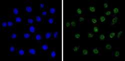
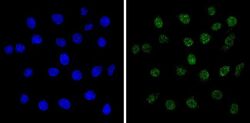
- Immunofluorescent analysis of Methyl CpG Binding Protein 2 (green) showing staining in the nucleus of C6 cells (right) compared to a negative control without primary antibody (left). Formalin-fixed cells were permeabilized with 0.1% Triton X-100 in TBS for 5-10 minutes and blocked with 3% BSA-PBS for 30 minutes at room temperature. Cells were probed with a Methyl CpG Binding Protein 2 polyclonal antibody (Product # PA1-887) in 3% BSA-PBS at a dilution of 1:200 and incubated overnight at 4ºC in a humidified chamber. Cells were washed with PBST and incubated with a DyLight-conjugated secondary antibody in PBS at room temperature in the dark. Nuclei were stained with Hoechst or DAPI (blue). Images were taken at a magnification of 60x.
- Submitted by
- Invitrogen Antibodies (provider)
- Main image
- Experimental details
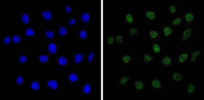
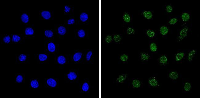
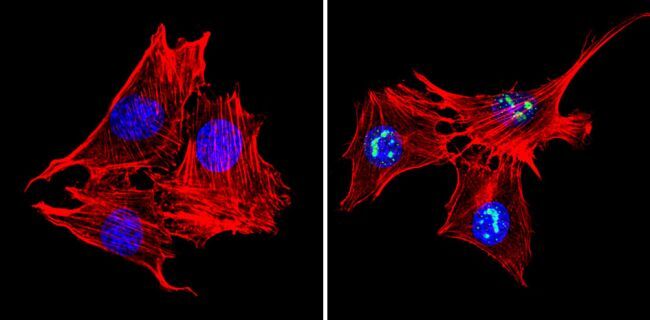
- Immunofluorescent analysis of Methyl CpG Binding Protein 2 (green) showing staining in the nucleus of C2C12 cells (right) compared to a negative control without primary antibody (left). Formalin-fixed cells were permeabilized with 0.1% Triton X-100 in TBS for 5-10 minutes and blocked with 3% BSA-PBS for 30 minutes at room temperature. Cells were probed with a Methyl CpG Binding Protein 2 polyclonal antibody (Product # PA1-887) in 3% BSA-PBS at a dilution of 1:200 and incubated overnight at 4ºC in a humidified chamber. Cells were washed with PBST and incubated with a DyLight-conjugated secondary antibody in PBS at room temperature in the dark. Actin was stained using Alexa Fluor 554 (red) and nuclei were stained with Hoechst or DAPI (blue). Images were taken at a magnification of 60x.

- Submitted by
- Invitrogen Antibodies (provider)
- Main image
- Experimental details
- Immunofluorescent analysis of Methyl CpG Binding Protein 2 (green) showing staining in the nucleus of C6 cells (right) compared to a negative control without primary antibody (left). Formalin-fixed cells were permeabilized with 0.1% Triton X-100 in TBS for 5-10 minutes and blocked with 3% BSA-PBS for 30 minutes at room temperature. Cells were probed with a Methyl CpG Binding Protein 2 polyclonal antibody (Product # PA1-887) in 3% BSA-PBS at a dilution of 1:200 and incubated overnight at 4ºC in a humidified chamber. Cells were washed with PBST and incubated with a DyLight-conjugated secondary antibody in PBS at room temperature in the dark. Nuclei were stained with Hoechst or DAPI (blue). Images were taken at a magnification of 60x.
- Submitted by
- Invitrogen Antibodies (provider)
- Main image

- Experimental details
- Immunofluorescent analysis of Methyl CpG Binding Protein 2 (green) showing staining in the nucleus of C2C12 cells (right) compared to a negative control without primary antibody (left). Formalin-fixed cells were permeabilized with 0.1% Triton X-100 in TBS for 5-10 minutes and blocked with 3% BSA-PBS for 30 minutes at room temperature. Cells were probed with a Methyl CpG Binding Protein 2 polyclonal antibody (Product # PA1-887) in 3% BSA-PBS at a dilution of 1:200 and incubated overnight at 4ºC in a humidified chamber. Cells were washed with PBST and incubated with a DyLight-conjugated secondary antibody in PBS at room temperature in the dark. Actin was stained using Alexa Fluor 554 (red) and nuclei were stained with Hoechst or DAPI (blue). Images were taken at a magnification of 60x.
- Submitted by
- Invitrogen Antibodies (provider)
- Main image
- Experimental details
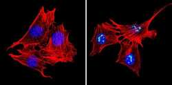
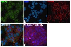
- Immunofluorescence analysis of Mecp2 was performed using 70% confluent log phase MCF7 cells. The cells were fixed with 4% paraformaldehyde for 10 minutes, permeabilized with 0.1% Triton™ X-100 for 15 minutes, and blocked with 2% BSA for 1 hour at room temperature. The cells were labeled with MECP2 Polyclonal Antibody (Product # PA1-887) at 1:200 in 0.1% BSA, incubated at 4 degree celsius overnight and then labeled with Donkey anti-Rabbit IgG (H+L) Highly Cross-Adsorbed Secondary Antibody, Alexa Fluor™ Plus 488 (Product # A32790, 1:2000), for 45 minutes at room temperature (Panel a: Green). Nuclei (Panel b:Blue) were stained with ProLong™ Diamond Antifade Mountant with DAPI (Product # P36962). F-actin (Panel c: Red) was stained with Rhodamine Phalloidin (Product # R415, 1:300). Panel d represents the merged image showing predominant nuclear localization. Panel e represents control cells with no primary antibody to assess background. The images were captured at 40X magnification.

Supportive validation
- Submitted by
- Invitrogen Antibodies (provider)
- Main image
- Experimental details
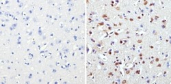
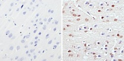
- Immunohistochemistry analysis of Methyl CpG Binding Protein 2 showing staining in the nucleus of paraffin-embedded mouse brain tissue (right) compared to a negative control without primary antibody (left). To expose target proteins, antigen retrieval was performed using 10mM sodium citrate (pH 6.0), microwaved for 8-15 min. Following antigen retrieval, tissues were blocked in 3% H2O2-methanol for 15 min at room temperature, washed with ddH2O and PBS, and then probed with a Methyl CpG Binding Protein 2 polyclonal antibody (Product # PA1-887) diluted in 3% BSA-PBS at a dilution of 1:500 overnight at 4°C in a humidified chamber. Tissues were washed extensively in PBST and detection was performed using an HRP-conjugated secondary antibody followed by colorimetric detection using a DAB kit. Tissues were counterstained with hematoxylin and dehydrated with ethanol and xylene to prep for mounting.
- Submitted by
- Invitrogen Antibodies (provider)
- Main image
- Experimental details
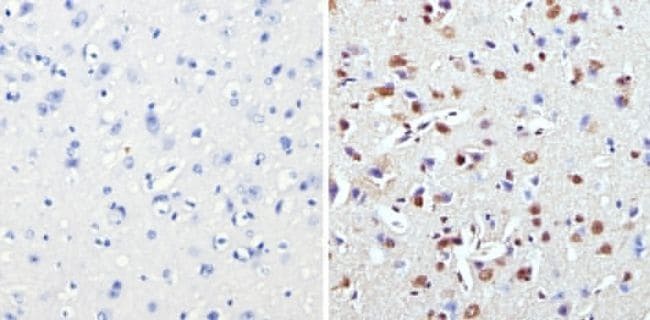
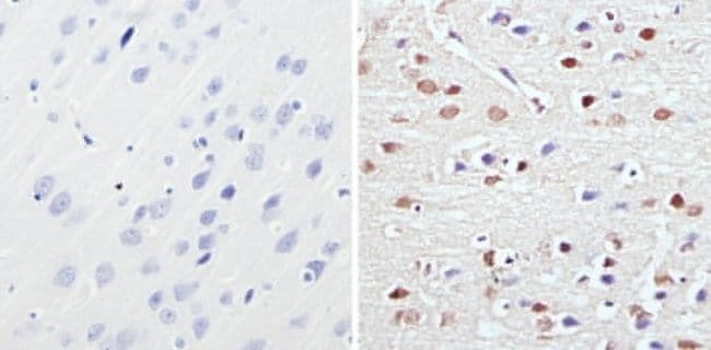
- Immunohistochemistry analysis of Methyl CpG Binding Protein 2 showing staining in the nucleus of paraffin-embedded rat brain tissue (right) compared to a negative control without primary antibody (left). To expose target proteins, antigen retrieval was performed using 10mM sodium citrate (pH 6.0), microwaved for 8-15 min. Following antigen retrieval, tissues were blocked in 3% H2O2-methanol for 15 min at room temperature, washed with ddH2O and PBS, and then probed with a Methyl CpG Binding Protein 2 polyclonal antibody (Product # PA1-887) diluted in 3% BSA-PBS at a dilution of 1:1000 overnight at 4°C in a humidified chamber. Tissues were washed extensively in PBST and detection was performed using an HRP-conjugated secondary antibody followed by colorimetric detection using a DAB kit. Tissues were counterstained with hematoxylin and dehydrated with ethanol and xylene to prep for mounting.
Supportive validation
- Submitted by
- Invitrogen Antibodies (provider)
- Main image

- Experimental details
- NULL
- Submitted by
- Invitrogen Antibodies (provider)
- Main image
- Experimental details
- NULL
- Submitted by
- Invitrogen Antibodies (provider)
- Main image
- Experimental details
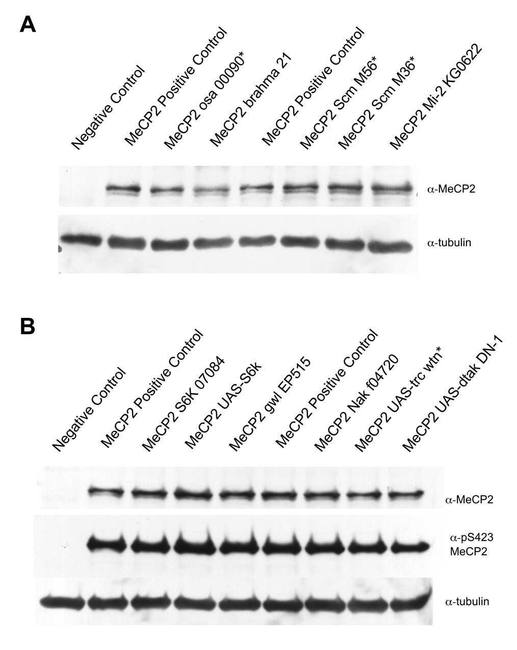
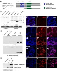
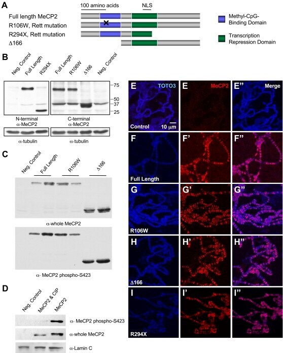
- Figure 1 MeCP2 alleles used to generate transgenic Drosophila : protein expression, phosphorylation at serine 423, and association with polytene chromosomes. A. Four MECP2 alleles were cloned into pUAST to generate transgenic flies. The methyl-CpG-binding domain (MBD) is represented by blue boxes and the transcription repression domain (TRD) is represented by green boxes. The nuclear localization signal (NLS) falls within the TRD. B. Western blot analysis demonstrates expression of each of the alleles when driven by GMR-Gal4 . Two distinct MeCP2 antibodies were utilized in order to recognize each allele to confirm that a deletion removed an epitope region. C. Immunoblot with a phospho-specific antibody shows phosphorylation in the three alleles retaining amino acid S423. D. Immunoblot with the phospho-specific MeCP2 S423 antibody in negative control, extracts from MeCP2 expressing flies when treated with calf intestinal phosphatase and untreated MeCP2 extracts. The treated samples fail to produce a band with the phospho-specific antibody, but demonstrate MeCP2 expression with the whole MeCP2 antibody (E-I""). Immunoflourescence of squashed polytene chromosomes dissected from 3rd instar larvae raised at 25degC. Control larvae do not have MeCP2 immunoreactivity (E-E""). All MeCP2 alleles demonstrate accumulation of the MeCP2 protein in banded pattern along the polytene chromosomes (F-I"").
- Submitted by
- Invitrogen Antibodies (provider)
- Main image

- Experimental details
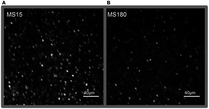
- Figure 7 Representative photomicrographs of immunolabeling for MeCP2 in the NAc core . (A) MS15, (B) MS180. Scale bar represents 40 mum.
- Submitted by
- Invitrogen Antibodies (provider)
- Main image
- Experimental details
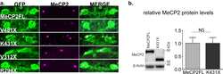
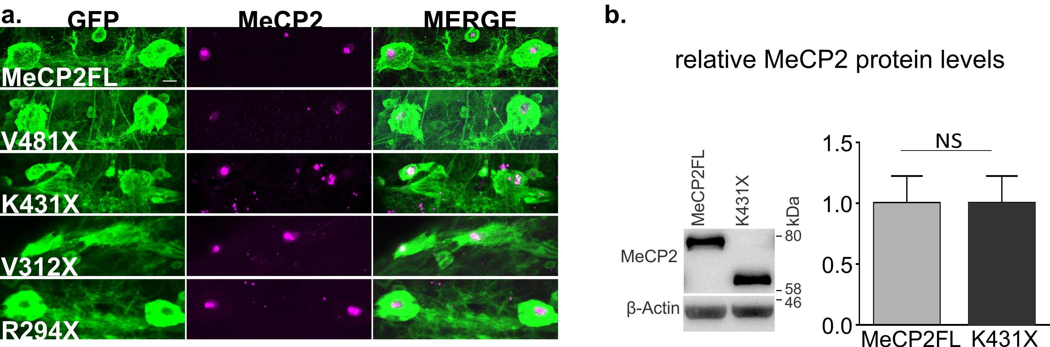
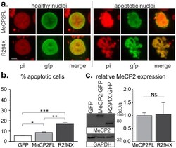
- Fig 2 R294X transfection promotes cell death in mammalian cell culture to a higher degree than MECP2FL . a. Representative images of HEK293T cells transfected with GFP tagged MECP2FL or R294X . Examples of healthy transfected cells are at left, while the condensed and/or fragmented nuclei at right were counted as apoptotic. b. Quantification of apoptotic cells following transfection of MECP2FL : GFP , R294X : GFP , or GFP control. n = 6 independent transfections/group, with > 400 cells counted for each independent transfection. Percentage data was transformed using the arcsine square root function to meet the assumptions for a one way ANOVA (F(2,15) = 46.81, p < .0001). * p < .05, ** p < .01, *** p < .001 Tukey post-hoc test. c. Relative MECP2 expression 24 hours following transfection into HEK293T cells. No differences were observed in relative protein levels at 24 or 48 hours post-transfection (Mann-Whitney U test). Scale bar depicts 5mum. pi = propidium iodide, NS = not significant.