Explore
 Validate
 Learn
Learn Western blot
 Immunocytochemistry
Antibody data
- Antibody Data
- Antigen structure
- References [1]
- Comments [0]
- Validations
- Immunocytochemistry [1]
- Immunohistochemistry [5]
- Other assay [1]
Submit
Validation data
Reference
Comment
Report error
- Product number
- PA5-99462 - Provider product page

- Provider
- Invitrogen Antibodies
- Product name
- CDKN2C Polyclonal Antibody
- Antibody type
- Polyclonal
- Antigen
- Synthetic peptide
- Description
- Antibody detects endogenous levels of total p18 INK4c.
- Reactivity
- Human, Mouse, Rat
- Host
- Rabbit
- Isotype
- IgG
- Vial size
- 100 μL
- Concentration
- 1 mg/mL
- Storage
- -20°C
Submitted references Bone morphogenetic protein 4 rescues the bone regenerative potential of old muscle-derived stem cells via regulation of cell cycle inhibitors.
Cheng H, Gao X, Huard M, Lu A, Ruzbarsky JJ, Amra S, Wang B, Huard J
Stem cell research & therapy 2022 Jul 30;13(1):385
Stem cell research & therapy 2022 Jul 30;13(1):385
No comments: Submit comment
Supportive validation
- Submitted by
- Invitrogen Antibodies (provider)
- Main image
- Experimental details
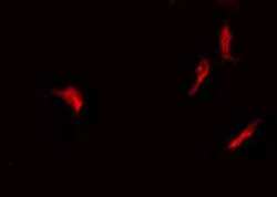
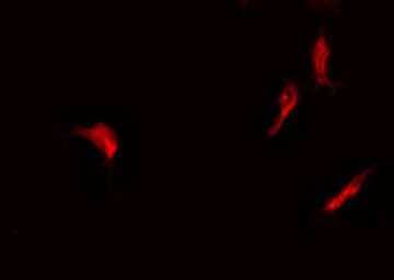
- Immunofluorescent analysis of CDKN2C in HeLa cells. Samples were fixed with paraformaldehyde, permeabilized with 0.1% Triton X-100, blocked with 10% serum (45 min at 25°C) incubated with CDKN2C polyclonal antibody (Product # PA5-99462) using a dilution of 1:200 (1 hr, 37°C), and followed by goat anti-rabbit IgG Alexa Fluor 594 at a dilution of 1:600.
Supportive validation
- Submitted by
- Invitrogen Antibodies (provider)
- Main image
- Experimental details
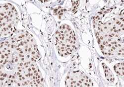
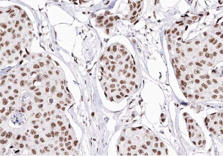
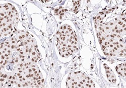
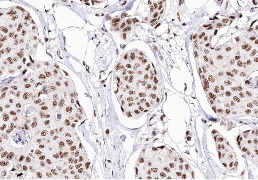
- Immunohistochemistry analysis of paraffin-embedded CDKN2C in human Breast cancer tissue sections. Antigen retrieval was performed using citrate buffer. Samples were blocked with blocking buffer (1.5 hr, 22°C), incubated with CDKN2C polyclonal antibody (Product # PA5-99462) using a dilution of 1:200 (1.5 hr, 22°C), followed by HRP conjugated goat anti-rabbit.
- Submitted by
- Invitrogen Antibodies (provider)
- Main image

- Experimental details
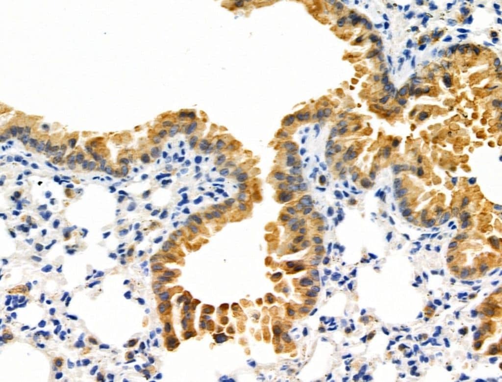
- Immunohistochemistry analysis of CDKN2C in mouse lung tissue. The sample was formaldehyde fixed and a heat mediated antigen retrieval step in citrate buffer was performed. Samples were incubated with CDKN2C polyclonal antibody (Product # PA5-99462) using a dilution of 1:100 (4°C overnight) followed by HRP conjugated anti-Rabbit secondary antibody.

- Submitted by
- Invitrogen Antibodies (provider)
- Main image
- Experimental details
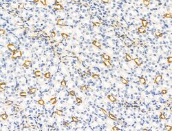
- Immunohistochemistry analysis of CDKN2C in mouse kidney tissue. The sample was formaldehyde fixed and a heat mediated antigen retrieval step in citrate buffer was performed. Samples were incubated with CDKN2C polyclonal antibody (Product # PA5-99462) using a dilution of 1:100 (4°C overnight) followed by HRP conjugated anti-Rabbit secondary antibody.

- Submitted by
- Invitrogen Antibodies (provider)
- Main image

- Experimental details
- Immunohistochemistry analysis of CDKN2C in mouse lung tissue. The sample was formaldehyde fixed and a heat mediated antigen retrieval step in citrate buffer was performed. Samples were incubated with CDKN2C polyclonal antibody (Product # PA5-99462) using a dilution of 1:100 (4°C overnight) followed by HRP conjugated anti-Rabbit secondary antibody.
- Submitted by
- Invitrogen Antibodies (provider)
- Main image
- Experimental details
- Immunohistochemistry analysis of paraffin-embedded CDKN2C in human Breast cancer tissue sections. Antigen retrieval was performed using citrate buffer. Samples were blocked with blocking buffer (1.5 hr, 22°C), incubated with CDKN2C polyclonal antibody (Product # PA5-99462) using a dilution of 1:200 (1.5 hr, 22°C), followed by HRP conjugated goat anti-rabbit.
Supportive validation
- Submitted by
- Invitrogen Antibodies (provider)
- Main image

- Experimental details
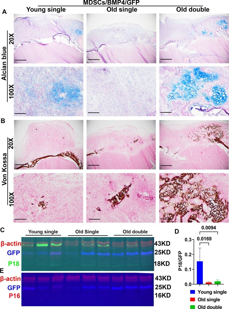
- Bone regeneration at day 10 after in vivo transplantation of young and old BMP4/GFP-transduced MDSCs. A Alcian blue staining. Chondrocytes and cartilage stained blue. 20X magnification shows the entire defect area. Transplanted cells proliferated and formed thick cell layers on top of the critical sized defect. Old MDSCs with double transduction showed early endochondral bone formation at day 10 after transplantation with obvious chondrogenic nodule in blue as did young MDSCs. Scale bars = 1 mm for 20x and 200 um for 100x, respectively. B Von Kossa Staining. Bone stained brown-black. Both young and old BMP4/GFP transduced MDSCs showed mineralization, old MDSCs with double BMP4/GFP transduction showed more mineralization than single transduction. These findings indicate that higher levels of BMP4 reversed the impairment of bone regeneration of old MDSCs. Scale bars = 1 mm for 20x and 200 um for 100x, respectively. C Fluorescent western blot for P18 (green) and GFP (blue) with beta-actin (red) as loading control. There is a green non-specific band in the P18 channel merging with the beta-actin (red) channel. D Quantification of P18 expression relative to GFP showed old MDSCs/BMP4/GFP expressed less P18 compared to young MDSCs/BMP4/GFP, while increased BMP4 expression by double transduction slightly increased P18 expression. E Western blot of P16 (Red) and GFP (blue) and loading control beta-actin (red) at day 10 after cell transplantation. P16 is not detectable by western blot