Explore
 Validate
 Learn
Learn Western blot
 Immunocytochemistry
Antibody data
- Antibody Data
- Antigen structure
- References [0]
- Comments [0]
- Validations
- Immunocytochemistry [4]
- Immunohistochemistry [5]
- Flow cytometry [1]
Submit
Validation data
Reference
Comment
Report error
- Product number
- MA5-52461 - Provider product page

- Provider
- Invitrogen Antibodies
- Product name
- RAB1A Recombinant Rabbit Monoclonal Antibody (PSH03-62)
- Antibody type
- Monoclonal
- Antigen
- Synthetic peptide
- Description
- Positive Control: U-87 MG cell lysate, U-2 OS cell lysate, A549 cell lysate, HCT 116 cell lysate, NIH/3T3 cell lysate, C2C12 cell lysate, C6 cell lysate, mouse brain tissue lysate, mouse liver tissue lysate, rat brain tissue lysate, rat liver tissue lysate, human kidney tissue, mouse brain tissue, mouse kidney tissue, rat brain tissue, rat kidney tissue, U-87 MG, C2C12, C6. Subcellular Location: Golgi apparatus, Endoplasmic reticulum, Early endosome, Cytoplasm, cytosol, Membrane, Melanosome. Predicted band size: 23 kDa.
- Reactivity
- Human, Mouse, Rat
- Host
- Rabbit
- Isotype
- IgG
- Antibody clone number
- PSH03-62
- Vial size
- 100 μL
- Concentration
- 1 mg/mL
- Storage
- Store at 4°C short term. For long term storage, store at -20°C, avoiding freeze/thaw cycles.
No comments: Submit comment
Supportive validation
- Submitted by
- Invitrogen Antibodies (provider)
- Main image
- Experimental details
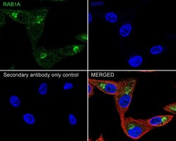
- Immunofluorescence analysis of RAB1A using U-87 MG cells. The cells were fixed 4% paraformaldehyde for 20 minutes, permeabilized with 0.1% Triton™ X-100 in PBS for 5 minutes, and blocked with 1% BSA in 10% negative goat serum for 1 hour at room temperature. The cells were labeled with RAB1A Recombinant Rabbit Monoclonal Antibody (PSH03-62) (Product # MA5-52461) (green) and beta tubulin (red) at 1:100 dilution in in 1% BSA in PBST overnight at 4 degrees Celsius and then with iFluor™ 488 Goat Anti-Rabbit IgG H&L and iFluor™ 594 Goat Anti-Mouse IgG H&L secondaries antibodies, respectively, at 1:1,000 dilution for 1 hour at room temperature. Nuclei were stained with DAPI (blue). The images were captured at 200X magnification.
- Submitted by
- Invitrogen Antibodies (provider)
- Main image
- Experimental details
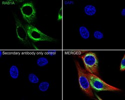
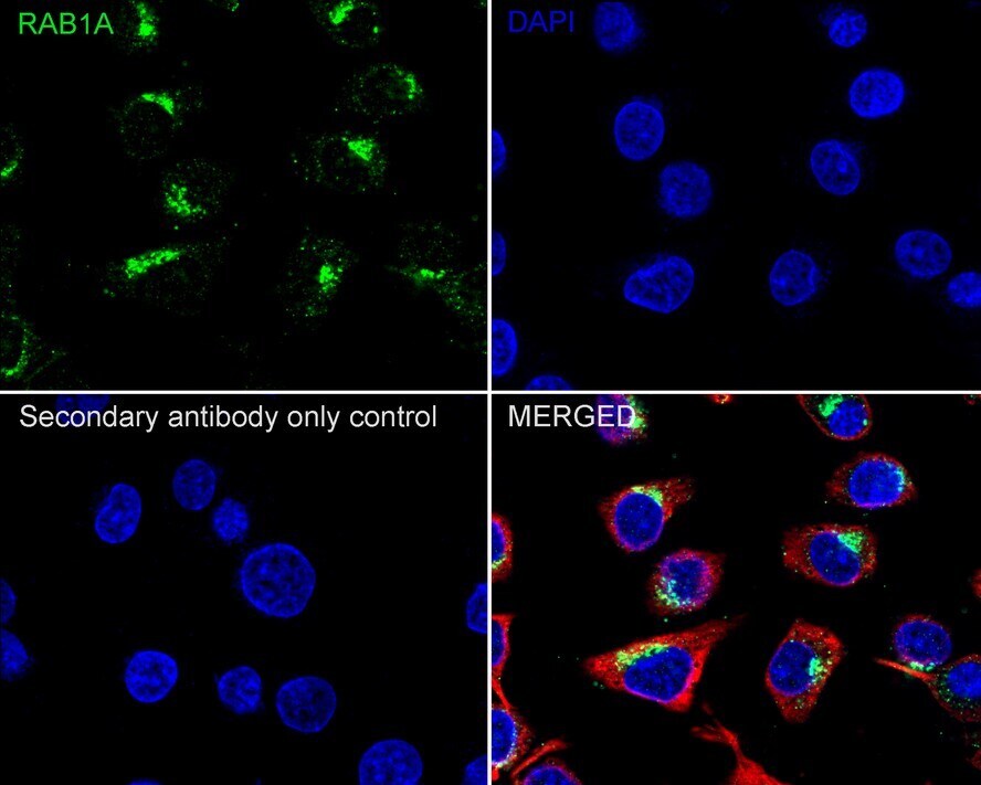
- Immunofluorescence analysis of RAB1A using C2C12 cells. The cells were fixed 4% paraformaldehyde for 20 minutes, permeabilized with 0.1% Triton™ X-100 in PBS for 5 minutes, and blocked with 1% BSA in 10% negative goat serum for 1 hour at room temperature. The cells were labeled with RAB1A Recombinant Rabbit Monoclonal Antibody (PSH03-62) (Product # MA5-52461) (green) and beta tubulin (red) at 1:100 dilution in in 1% BSA in PBST overnight at 4 degrees Celsius and then with iFluor™ 488 Goat Anti-Rabbit IgG H&L and iFluor™ 594 Goat Anti-Mouse IgG H&L secondaries antibodies, respectively, at 1:1,000 dilution for 1 hour at room temperature. Nuclei were stained with DAPI (blue). The images were captured at 200X magnification.

- Submitted by
- Invitrogen Antibodies (provider)
- Main image
- Experimental details
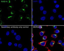
- Immunofluorescence analysis of RAB1A using C6 cells. The cells were fixed 4% paraformaldehyde for 20 minutes, permeabilized with 0.1% Triton™ X-100 in PBS for 5 minutes, and blocked with 1% BSA in 10% negative goat serum for 1 hour at room temperature. The cells were labeled with RAB1A Recombinant Rabbit Monoclonal Antibody (PSH03-62) (Product # MA5-52461) (green) and beta tubulin (red) at 1:100 dilution in in 1% BSA in PBST overnight at 4 degrees Celsius and then with iFluor™ 488 Goat Anti-Rabbit IgG H&L and iFluor™ 594 Goat Anti-Mouse IgG H&L secondaries antibodies, respectively, at 1:1,000 dilution for 1 hour at room temperature. Nuclei were stained with DAPI (blue). The images were captured at 200X magnification.
- Submitted by
- Invitrogen Antibodies (provider)
- Main image
- Experimental details
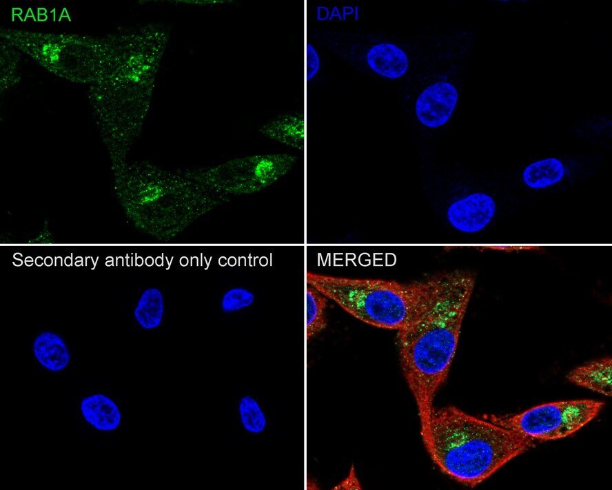
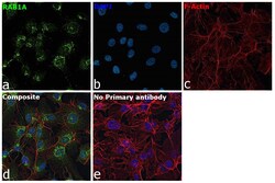
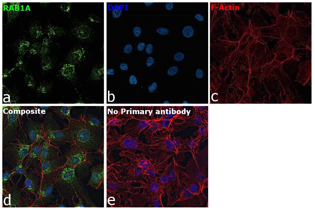
- Immunofluorescence analysis of RAB1A was performed using 70% confluent log phase Hep G2 cells. The cells were fixed with 4% paraformaldehyde for 10 minutes, permeabilized with 0.1% Triton™ X-100 for 15 minutes, and blocked with 2% BSA for 1 hour at room temperature. The cells were labeled with RAB1A Recombinant Rabbit Monoclonal Antibody (PSH03-62) (Product # MA5-52461) at 1:100 dilution in 0.1% BSA, incubated at 4 degree Celsius overnight and then labeled with Donkey anti-Rabbit IgG (H+L) Highly Cross-Adsorbed Secondary Antibody, Alexa Fluor Plus 488 (Product # A32790), (1:2,000 dilution), for 45 minutes at room temperature (Panel a: Green). Nuclei (Panel b:Blue) were stained with ProLong™ Diamond Antifade Mountant with DAPI (Product # P36962). F-actin (Panel c: Red) was stained with Rhodamine Phalloidin (Product # R415, 1:300 dilution). Panel d represents the merged image showing predominantly golgi and endoplasmic reticulum localization. Panel e represents control cells with no primary antibody to assess background. The images were captured at 40X magnification.
Supportive validation
- Submitted by
- Invitrogen Antibodies (provider)
- Main image
- Experimental details
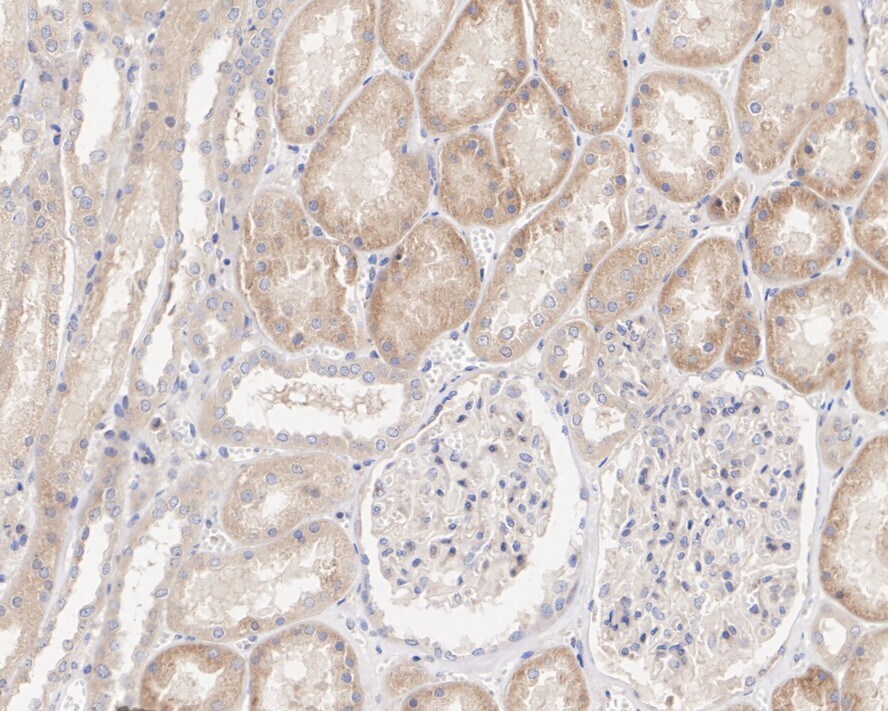
- Immunohistochemical analysis of RAB1A on formalin-fixed paraffin-embedded rat kidney tissue. The section was pre-treated using heat mediated antigen retrieval with Tris-EDTA buffer (pH 9.0) for 20 minutes. The tissue was blocked in 1% BSA for 20 minutes at room temperature, then probed with RAB1A Recombinant Rabbit Monoclonal Antibody (PSH03-62) (Product # MA5-52461) at 1:200 dilution for 1 hour at room temperature. HRP conjugated compact polymer system and DAB chromogen were used as the detection system, followed by counterstaining with hematoxylin. The slide was mounted with DPX and the image was captured at 200X magnification.

- Submitted by
- Invitrogen Antibodies (provider)
- Main image

- Experimental details
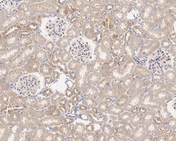
- Immunohistochemical analysis of RAB1A on formalin-fixed paraffin-embedded human kidney tissue. The section was pre-treated using heat mediated antigen retrieval with Tris-EDTA buffer (pH 9.0) for 20 minutes. The tissue was blocked in 1% BSA for 20 minutes at room temperature, then probed with RAB1A Recombinant Rabbit Monoclonal Antibody (PSH03-62) (Product # MA5-52461) at 1:200 dilution for 1 hour at room temperature. HRP conjugated compact polymer system and DAB chromogen were used as the detection system, followed by counterstaining with hematoxylin. The slide was mounted with DPX and the image was captured at 200X magnification.
- Submitted by
- Invitrogen Antibodies (provider)
- Main image
- Experimental details
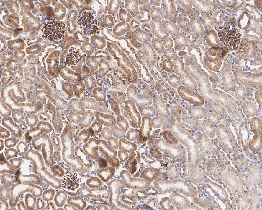
- Immunohistochemical analysis of RAB1A on formalin-fixed paraffin-embedded mouse kidney tissue. The section was pre-treated using heat mediated antigen retrieval with Tris-EDTA buffer (pH 9.0) for 20 minutes. The tissue was blocked in 1% BSA for 20 minutes at room temperature, then probed with RAB1A Recombinant Rabbit Monoclonal Antibody (PSH03-62) (Product # MA5-52461) at 1:200 dilution for 1 hour at room temperature. HRP conjugated compact polymer system and DAB chromogen were used as the detection system, followed by counterstaining with hematoxylin. The slide was mounted with DPX and the image was captured at 200X magnification.
- Submitted by
- Invitrogen Antibodies (provider)
- Main image
- Experimental details
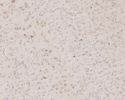
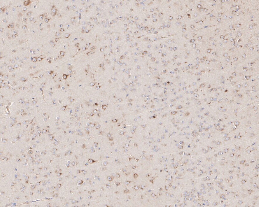
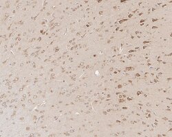
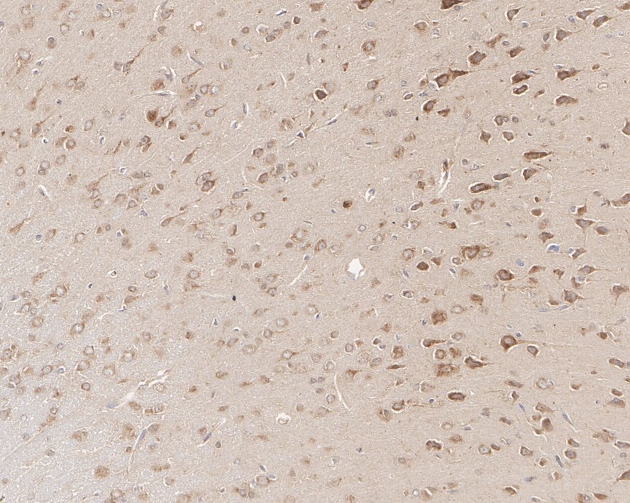
- Immunohistochemical analysis of RAB1A on formalin-fixed paraffin-embedded mouse brain tissue. The section was pre-treated using heat mediated antigen retrieval with Tris-EDTA buffer (pH 9.0) for 20 minutes. The tissue was blocked in 1% BSA for 20 minutes at room temperature, then probed with RAB1A Recombinant Rabbit Monoclonal Antibody (PSH03-62) (Product # MA5-52461) at 1:200 dilution for 1 hour at room temperature. HRP conjugated compact polymer system and DAB chromogen were used as the detection system, followed by counterstaining with hematoxylin. The slide was mounted with DPX and the image was captured at 200X magnification.
- Submitted by
- Invitrogen Antibodies (provider)
- Main image
- Experimental details
- Immunohistochemical analysis of RAB1A on formalin-fixed paraffin-embedded rat brain tissue. The section was pre-treated using heat mediated antigen retrieval with Tris-EDTA buffer (pH 9.0) for 20 minutes. The tissue was blocked in 1% BSA for 20 minutes at room temperature, then probed with RAB1A Recombinant Rabbit Monoclonal Antibody (PSH03-62) (Product # MA5-52461) at 1:200 dilution for 1 hour at room temperature. HRP conjugated compact polymer system and DAB chromogen were used as the detection system, followed by counterstaining with hematoxylin. The slide was mounted with DPX and the image was captured at 200X magnification.
Supportive validation
- Submitted by
- Invitrogen Antibodies (provider)
- Main image
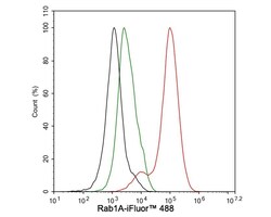
- Experimental details
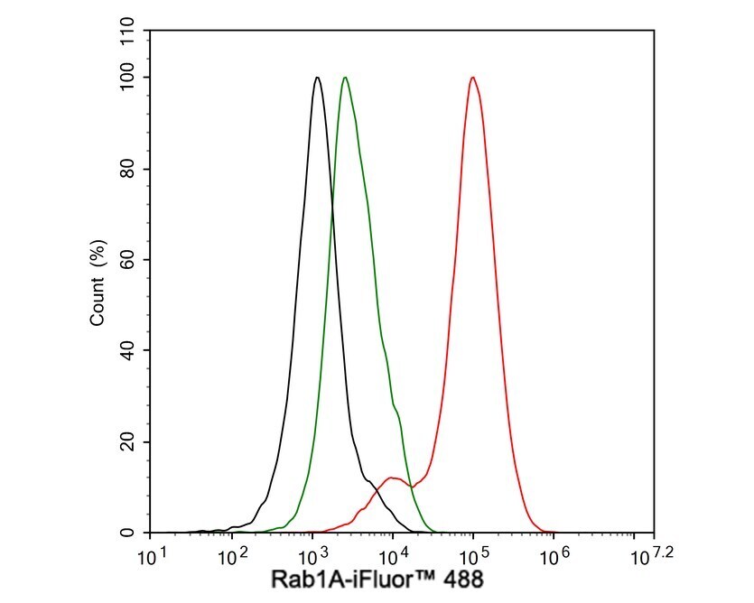
- U-87 MG cells were fixed and permeabilized and then stained with RAB1A Recombinant Rabbit Monoclonal Antibody (PSH03-62) (Product # MA5-52461) at 1 μg/mL (red) or Rabbit IgG Isotype Control (green). After incubation of the primary antibody at 4 degrees Celsius for an hour, the cells were stained with a iFluor™ 488 conjugate-Goat anti-Rabbit IgG Secondary antibody at 1:1,000 dilution for 30 minutes at 4 degrees Celsius. Unlabelled sample was used as a control (cells without incubation with primary antibody; black).