Explore
 Validate
 Learn
Learn Western blot
 Immunocytochemistry
 Immunohistochemistry
Antibody data
- Antibody Data
- Antigen structure
- References [0]
- Comments [0]
- Validations
- Western blot [3]
- Chromatin Immunoprecipitation [1]
- Other assay [3]
Submit
Validation data
Reference
Comment
Report error
- Product number
- PA5-19546 - Provider product page

- Provider
- Invitrogen Antibodies
- Product name
- Anti-SAM68 Polyclonal Antibody
- Antibody type
- Polyclonal
- Antigen
- Synthetic peptide
- Description
- Heat mediated antigen retrieval recommended prior to tissue staining. This antibody is predicted to react with chicken and dog based on sequence homology.
- Reactivity
- Human, Mouse, Rat
- Host
- Rabbit
- Isotype
- IgG
- Vial size
- 100 µg
- Concentration
- 1 mg/mL
- Storage
- Store at 4°C short term. For long term storage, store at -20°C, avoiding freeze/thaw cycles.
No comments: Submit comment
Supportive validation
- Submitted by
- Invitrogen Antibodies (provider)
- Main image
- Experimental details
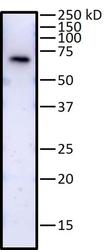
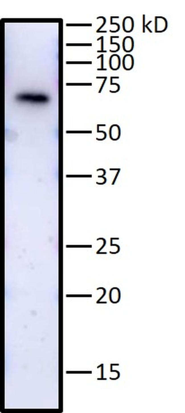
- Western blot analysis of SAM68 was performed by loading 10 µg of whole cell human BJ fibroblast protein lysate and run on a 4-12% BTE gel. Proteins were transferred to PVDF membrane. Membrane was blocked with Pierce™ Protein-Free T20 (PBS) Blocking Buffer (Product # 37573). SAM68 was detected at approximately 68 kDa using a SAM68 polyclonal antibody (Product # PA5-19546) at a dilution of 1:1000 followed by a 1:10,000 dilution of anti-rabbit HRP. Chemiluminescent detection was performed using SuperSignal West Pico PLUS substrate (Product # 34580). Data courtesy of Antibody Data Exchange Program.
- Submitted by
- Invitrogen Antibodies (provider)
- Main image
- Experimental details
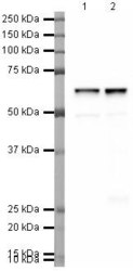
- Western blot analysis of HeLa Whole Cell Lysate using Product # PA5-19546, SAM68 primary antibody at a dilution of 1 µg/mL (lane 1). Staining of Jurkat Whole Cell Lysate at a dilution of 1 µg/mL (lane 2). Blot treated with a secondary HRP-conjugated Goat polyclonal anti-Rabbit antibody was used at a dilution of 1:3000.

- Submitted by
- Invitrogen Antibodies (provider)
- Main image
- Experimental details
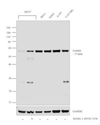
- Western blot analysis was performed on modified whole cell extracts (1% SDS) (30 µg lysate) of MCF7 (Lane 1), MCF7 treated with Insulin (1nM for 24hr) (Lane 2), HeLa (Lane 3), Jurkat (Lane 4), A-431 (Lane 5) and U-87 MG (Lane 6). The blot was probed with Anti-SAM68 Polyclonal Antibody (Product # PA5-19546, 1:1000 dilution) and detected by chemiluminescence using Goat anti-Rabbit IgG (H+L) Superclonal™ Secondary Antibody, HRP conjugate (Product # A27036, 0.25 µg/mL, 1:4000 dilution). A 57 kDa band corresponding to SAM68 was detected across the cell lines tested and the expression was also found to increase upon Insulin treatment in MCF7 cells.

Supportive validation
- Submitted by
- Invitrogen Antibodies (provider)
- Main image

- Experimental details
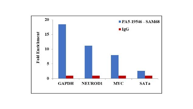
- Chromatin Immunoprecipitation (ChIP) assay of endogenous SAM68 protein using Anti-SAM68 Antibody: ChIP was performed using Anti-SAM68 Rabbit Polyclonal Antibody (Product # PA5-19546, 5 µg) on sheared chromatin from HeLa cells using the MAGnify ChIP System kit (Product # 49-2024). Normal Rabbit IgG was used as a negative IP control. The purified DNA was analyzed by qPCR using primers binding to GAPDH, NeuroD1 and cMYC transcriptional start sites, and SAT alpha satellite repeats. Data is presented as fold enrichment of the antibody signal versus the negative control IgG using the comparative CT method.
Supportive validation
- Submitted by
- Invitrogen Antibodies (provider)
- Main image
- Experimental details
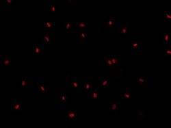
- Immunofluorescence analysis of SAM68 in human fibroblasts. Cells were fixed with 2% PFA in PBS and permeabilized with 0.1% Triton X-100 in PBS. Cells were blocked with 3% Donkey serum in PBS followed by staining with a SAM68 polyclonal antibody (Product # PA5-19546) at a dilution of 1:200. Cells washed extensively followed by an incubation with anti-rabbit Alexa Fluor 594 (Product # A-21207) at a dilution of 1:500 (red). Data courtesy of Antibody Data Exchange Program.
- Submitted by
- Invitrogen Antibodies (provider)
- Main image

- Experimental details
- Immunofluorescent staining of HEK293 cells using Product # PA5-19546, anti-SAM68 antibody. The cells were fixed with methanol (100%) for 5 minutes, permabilised with TBS-T (20mins), BSA (1%), normal goat serum (10%) and glycine (0.3 M) in 0.1% PBS-Tween for 1 hour and exposed to the primary antibody at a concentration of 1 µg/mL for 1 hour at room temp. The secondary antibody was a 448 fluorescence conjugated Goat anti-rabbit IgG (green) at a dilution of 1:1000. A WGA- 594 fluorescent conjugated stain was used to label plasma membranes (red) and the nuclei stain was DAPI (blue).
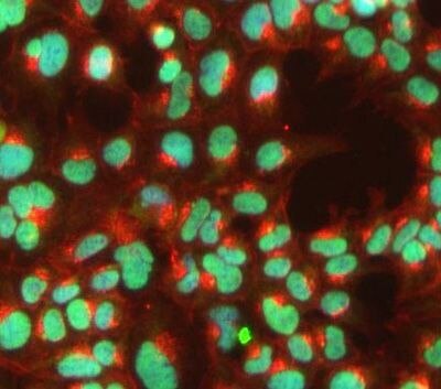
- Submitted by
- Invitrogen Antibodies (provider)
- Main image

- Experimental details
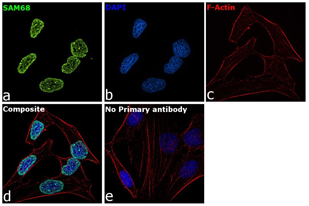
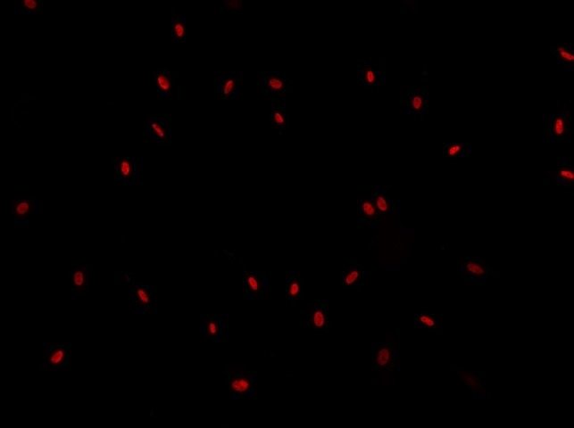
- Immunofluorescence analysis of SAM68 was performed using 70% confluent log phase HeLa cells. The cells were fixed with 4% paraformaldehyde for 10 minutes, permeabilized with 0.1% Triton™ X-100 for 15 minutes, and blocked with 1% BSA for 1 hour at room temperature. The cells were labeled with SAM68 Polyclonal Antibody (Product # PA5-19546) at 1µg/mL in 0.1% BSA, incubated at 4 degree Celsius overnight and then labeled with Goat anti-Rabbit IgG (H+L) Superclonal™ Secondary Antibody, Alexa Fluor® 488 conjugate (Product # A27034) at a dilution of 1:2000 for 45 minutes at room temperature (Panel a: green). Nuclei (Panel b: blue) were stained with ProLong™ Diamond Antifade Mountant with DAPI (Product # P36962). F-actin (Panel c: red) was stained with Rhodamine Phalloidin (Product # R415). Panel d represents the merged image showing nuclear localization. Panel e represents control cells with no primary antibody to assess background. The images were captured at 60X magnification.