 Explore
 Validate
 Learn
Learn Western blot
 Immunocytochemistry
Antibody data
- Antibody Data
- Antigen structure
- References [1]
- Comments [0]
- Validations
- Immunocytochemistry [1]
- Immunohistochemistry [1]
- Other assay [1]
Submit
Validation data
Reference
Comment
Report error
- Product number
- PA1-41220 - Provider product page

- Provider
- Invitrogen Antibodies
- Product name
- Nogo-A/B Polyclonal Antibody
- Antibody type
- Polyclonal
- Antigen
- Synthetic peptide
- Description
- Suggested positive control: human, mouse or rat brain.
- Reactivity
- Human, Mouse, Rat, Rabbit
- Host
- Rabbit
- Isotype
- IgG
- Vial size
- 200 μL
- Concentration
- 1.0 mg/mL
- Storage
- Store at 4°C short term. For long term storage, store at -20°C, avoiding freeze/thaw cycles.
Submitted references REEP5 depletion causes sarco-endoplasmic reticulum vacuolization and cardiac functional defects.
Lee SH, Hadipour-Lakmehsari S, Murthy HR, Gibb N, Miyake T, Teng ACT, Cosme J, Yu JC, Moon M, Lim S, Wong V, Liu P, Billia F, Fernandez-Gonzalez R, Stagljar I, Sharma P, Kislinger T, Scott IC, Gramolini AO
Nature communications 2020 Feb 19;11(1):965
Nature communications 2020 Feb 19;11(1):965
No comments: Submit comment
Supportive validation
- Submitted by
- Invitrogen Antibodies (provider)
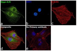
- Main image
- Experimental details
- Immunofluorescence analysis of Reticulon-4 was performed using 70% confluent log phase U-87 MG cells. The cells were fixed with 4% paraformaldehyde for 10 minutes, permeabilized with 0.1% Triton™ X-100 for 15 minutes, and blocked with 2% BSA for 45 minutes at room temperature. The cells were labeled with Nogo-A/B Polyclonal Antibody (Product # PA1-41220) at (1:100 dilution) in 0.1% BSA, incubated at 4-degree Celsius overnight, and labeled with Donkey anti-Rabbit IgG (H+L) Highly Cross-Adsorbed Secondary Antibody, Alexa Fluor Plus 488 (Product # A32790), (1:2000 dilution), for 45 minutes at room temperature (Panel a: Green). Nuclei (Panel b: Blue) were stained with ProLong™ Diamond Antifade Mountant with DAPI (Product # P36962). F-actin (Panel c: Red) was stained with Rhodamine Phalloidin (Product # R415, 1:300 dilution). Panel d represents the merged image showing cytoplasmic localization. Panel e represents control cells with no primary antibody to assess background. The images were captured at 60X magnification.
Supportive validation
- Submitted by
- Invitrogen Antibodies (provider)
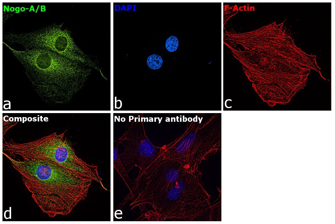
- Main image

- Experimental details
- Immunohistochemical analysis of Nogo-A/B in human testis. Samples were incubated in Nogo-A/B polyclonal antibody (Product # PA1-41220) using a dilution of 1:100.
Supportive validation
- Submitted by
- Invitrogen Antibodies (provider)
- Main image
- Experimental details
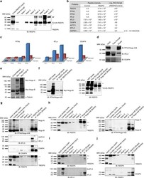
- Fig. 6 Mass spectrometry analysis identifies REEP5 interactions with known cardiac SR shaping proteins. a Immunoblot analysis of nickel-His purification of CnVA-REEP5 from transfected HEK293 cells shows stable expression of CnVA-REEP5 and successful immunoprecipitation of CnVA-REEP5 monomer (~34 kDa) and dimer (~68 kDa). b Identification of known SR/ER-shaping proteins as REEP5 interacting proteins by mass spectrometry analysis. Average precursor MS1 peak areas (peptide m/z signal) as defined by iBAQ (intensity based absolute quantification) are shown, n = 3 independent mass spectrometry runs. To calculate fold change, average values (n.d.--not detected) were inputted with a value of 10. RTN reticulon, ATL atlastin, CKAP4 cytoskeleton-associated protein 4, ACTC1 alpha cardiac muscle actin 1, MYL6 myosin light chain 6, GAPDH glyceraldehyde-3-phosphate dehydrogenase. Asterisks indicate a statistically significant p value in a Tukey's multiple comparison analysis where * p < 0.05. c RNASeq analysis of the RTN, ATL families of proteins, and CKAP4 in human fetal heart and adult heart tissue data using data from Human Protein Atlas. d Immunoblot analysis of nickel-His REEP5 immunoprecipitation lysates for RTN4/Nogo-A/B, ATL3, and CKAP4, n = 3. e HEK293 c e lls were transfected with myc-tagged Nogo-A and Nogo-B plasmids and detected with myc and alpha-tubulin antibodies. f Co-immunoprecipitation assay of cotransfected HEK293 cells with anti-REEP5 antibody (left panel) and anti-
