 Explore
 Validate
 Learn
Learn Western blot
Antibody data
- Antibody Data
- Antigen structure
- References [0]
- Comments [0]
- Validations
- Western blot [3]
- Immunocytochemistry [2]
- Immunohistochemistry [5]
Submit
Validation data
Reference
Comment
Report error
- Product number
- PA5-20367 - Provider product page

- Provider
- Invitrogen Antibodies
- Product name
- Nogo-A Polyclonal Antibody
- Antibody type
- Polyclonal
- Antigen
- Synthetic peptide
- Description
- Despite its predicted molecular weight, NogoA typically migrates at 180kDa in an SDS-PAGE. A suggested positive control is human brain tissue lysate. PA5-20367 can be used with blocking peptide PEP-0481.
- Reactivity
- Human, Mouse, Rat
- Host
- Rabbit
- Isotype
- IgG
- Vial size
- 100 µg
- Concentration
- 1 mg/mL
- Storage
- Maintain refrigerated at 2-8°C for up to 3 months. For long term storage store at -20°C
No comments: Submit comment
Supportive validation
- Submitted by
- Invitrogen Antibodies (provider)
- Main image

- Experimental details
- Western blot analysis of human brain tissue lysate using a NogoA polyclonal antibody (Product # PA5-20367) at (A) 0.5 and (B) 1 µg/mL.

- Submitted by
- Invitrogen Antibodies (provider)
- Main image

- Experimental details
- Western Blot analysis of NogoA in human brain tissue lysate with Nogo-A Polyclonal Antibody (Product # PA5-20367) at (A) 0.5 and (B) 1 µg/mL.

- Submitted by
- Invitrogen Antibodies (provider)
- Main image

- Experimental details
- Western Blot was performed using Anti-Nogo-A Polyclonal Antibody (Product # PA5-20367) and a 200 kDa band corresponding to Reticulon-4 (Nogo-A) was observed across all the cell lines and tissue lysates tested except for MCF7. A 50 kDa band corresponding to Nogo-B was also observed in all the cell lines tested. Membrane enriched extracts (30 µg lysate) of U-87 MG (Lane 1), SH-SY5Y (Lane 2), IMR-90 (Lane 3), Daudi (Lane 4), MCF7 (Lane 5), Mouse Brain (Lane 6), Rat Brain (Lane 7), Mouse Cerebellum (Lane 8), Rat Cerebellum (Lane 9), Mouse Heart (Lane 10), Rat Heart (Lane 11) were electrophoresed using NuPAGE™ 3-8% Tris-Acetate Protein Gel (Product # EA0378BOX). Resolved proteins were then transferred onto a Nitrocellulose membrane (Product # IB23001) by iBlot® 2 Dry Blotting System (Product # IB21001). The Blot was probed with the primary antibody (1 µg/mL) and detected by chemiluminescence with Goat anti-Rabbit IgG (H+L) Superclonal™ Recombinant Secondary Antibody, HRP (Product # A27036, 1:4000 dilution) using the iBright FL 1000 (Product # A32752). Chemiluminescent detection was performed using Novex® ECL Chemiluminescent Substrate Reagent Kit (Product # WP20005). Nogo-A expression was absent in the MCF7 cell line and lower in Mouse/Rat heart tissue lysates as compared to Mouse/Rat brain and cerebellum tissue lysates as reported in protein expression databases.

Supportive validation
- Submitted by
- Invitrogen Antibodies (provider)
- Main image

- Experimental details
- Immunofluorescent analysis of mouse brain cells using a NogoA polyclonal antibody (Product # PA5-20367) at a 20 µg/mL dilution.

- Submitted by
- Invitrogen Antibodies (provider)
- Main image

- Experimental details
- Immunofluorescence analysis of Reticulon-4 was performed using 70% confluent log phase U-87 MG cells. The cells were fixed with 4% paraformaldehyde for 10 minutes, permeabilized with 0.1% Triton™ X-100 for 15 minutes, and blocked with 2% BSA for 45 minutes at room temperature. The cells were labeled with Nogo-A Polyclonal Antibody (Product # PA5-20367) at 2.5 µg/mL in 0.1% BSA, incubated at 4-degree Celsius overnight, and then labeled with Donkey anti-Rabbit IgG (H+L) Highly Cross-Adsorbed Secondary Antibody, Alexa Fluor Plus 488 (Product # A32790), (1:2000 dilution), for 45 minutes at room temperature (Panel a: Green). Nuclei (Panel b:Blue) were stained with ProLong™ Diamond Antifade Mountant with DAPI (Product # P36962). F-actin (Panel c: Red) was stained with Rhodamine Phalloidin (Product # R415, 1:300 dilution). Panel d represents the merged image showing cytoplasmic localization. Panel e represents control cells with no primary antibody to assess the background. The images were captured at 60X magnification.

Supportive validation
- Submitted by
- Invitrogen Antibodies (provider)
- Main image
- Experimental details
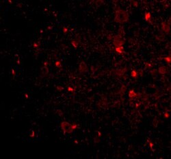
- Immunofluorescence of NogoA in Mouse Brain tissue with Nogo-A Polyclonal Antibody (Product # PA5-20367) at 20 µg/mL.

- Submitted by
- Invitrogen Antibodies (provider)
- Main image

- Experimental details
- Immunofluorescence of NogoA in mouse brain tissue with Nogo-A Polyclonal Antibody (Product # PA5-20367) at 20 µg/mL. Green: NogoA Red: Phylloidin staining Blue: DAPI staining

- Submitted by
- Invitrogen Antibodies (provider)
- Main image

- Experimental details
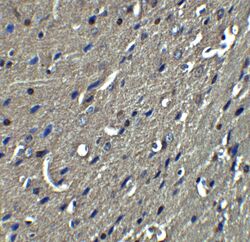
- Immunohistochemistry of NogoA in mouse brain tissue with Nogo-A Polyclonal Antibody (Product # PA5-20367) at 2.5 µg/mL.

- Submitted by
- Invitrogen Antibodies (provider)
- Main image
- Experimental details
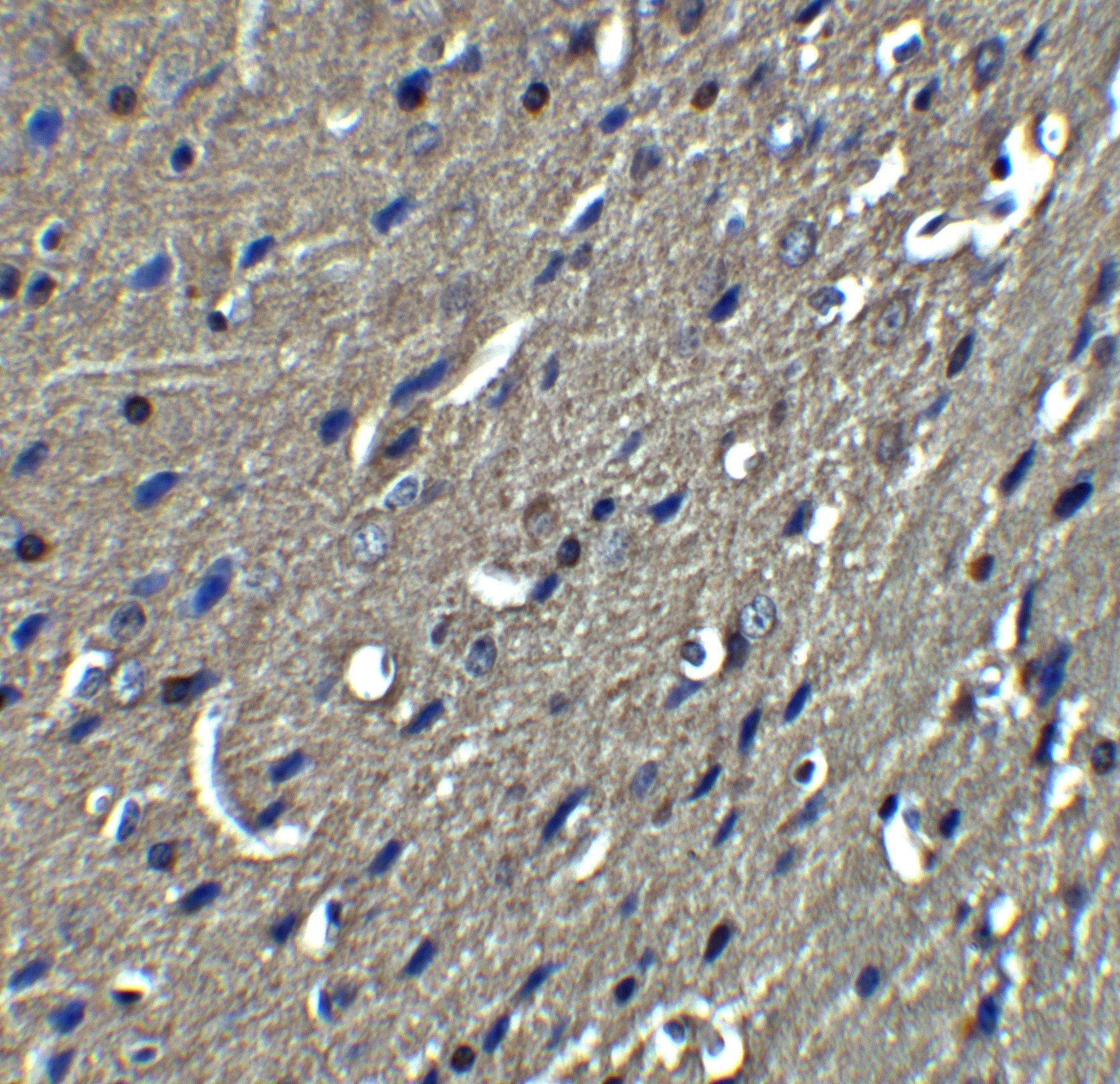
- Immunohistochemistry of NogoA in mouse brain tissue with Nogo-A Polyclonal Antibody (Product # PA5-20367) at 5 µg/mL.
- Submitted by
- Invitrogen Antibodies (provider)
- Main image
- Experimental details
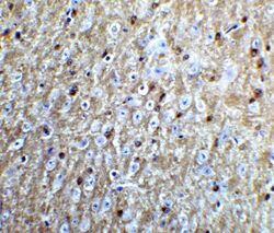
- Immunohistochemistry of NogoA in mouse brain tissue with Nogo-A Polyclonal Antibody (Product # PA5-20367) at 5 µg/mL.
