Explore
 Validate
 Learn
Learn Western blot
 Immunocytochemistry
Antibody data
- Antibody Data
- Antigen structure
- References [5]
- Comments [0]
- Validations
- Immunocytochemistry [6]
- Other assay [4]
Submit
Validation data
Reference
Comment
Report error
- Product number
- PA1-337 - Provider product page

- Provider
- Invitrogen Antibodies
- Product name
- SREBP1 Polyclonal Antibody
- Antibody type
- Polyclonal
- Antigen
- Synthetic peptide
- Description
- PA1-337 detects SREBP 1 in mouse and rat samples. PA1-337 has been successfully used in Western blot and ICC/IF procedures. By Western blot, this antibody detects an ~68 and 120 kDa protein representing SREBP 1 in mouse and rat liver samples as well as rat kidney samples. A predominent band at ~68 kDa (active cleaved site) is seen and a band at ~120 kDa (inactive precursor) may not be seen or it may be diminished. The PA1-337 immunogen is a synthetic peptide corresponding to residues M(32) L Q L I N N Q D S D F P G L F(47) of mouse SREBP 1. This sequence is identical in human and rat. PA1-337 immunizing peptide (Cat. # PEP-274) is available for use in neutralization and control experiments.
- Reactivity
- Human, Mouse, Rat
- Host
- Rabbit
- Isotype
- IgG
- Vial size
- 100 µg
- Concentration
- 1 mg/mL
- Storage
- -20°C, Avoid Freeze/Thaw Cycles
Submitted references Keratin 17 Is Required for Lipid Metabolism in Keratinocytes and Benefits Epidermal Permeability Barrier Homeostasis.
Gycyrrhizic acid alleviates atherosclerotic lesions in rats with diabetes mellitus.
Polygonatum sibiricum F. Delaroche polysaccharide ameliorates HFD‑induced mouse obesity via regulation of lipid metabolism and inflammatory response.
Long term N-acetylcysteine administration rescues liver steatosis via endoplasmic reticulum stress with unfolded protein response in mice.
Sterol regulatory element binding protein and dietary lipid regulation of fatty acid synthesis in the mammary epithelium.
Pang B, Zhu Z, Xiao C, Luo Y, Fang H, Bai Y, Sun Z, Ma J, Dang E, Wang G
Frontiers in cell and developmental biology 2021;9:779257
Frontiers in cell and developmental biology 2021;9:779257
Gycyrrhizic acid alleviates atherosclerotic lesions in rats with diabetes mellitus.
Zhao Y, Li W, Zhang D
Molecular medicine reports 2021 Nov;24(5)
Molecular medicine reports 2021 Nov;24(5)
Polygonatum sibiricum F. Delaroche polysaccharide ameliorates HFD‑induced mouse obesity via regulation of lipid metabolism and inflammatory response.
Liu B, Tang Y, Song Z, Ge J
Molecular medicine reports 2021 Jul;24(1)
Molecular medicine reports 2021 Jul;24(1)
Long term N-acetylcysteine administration rescues liver steatosis via endoplasmic reticulum stress with unfolded protein response in mice.
Tsai CC, Chen YJ, Yu HR, Huang LT, Tain YL, Lin IC, Sheen JM, Wang PW, Tiao MM
Lipids in health and disease 2020 May 25;19(1):105
Lipids in health and disease 2020 May 25;19(1):105
Sterol regulatory element binding protein and dietary lipid regulation of fatty acid synthesis in the mammary epithelium.
Rudolph MC, Monks J, Burns V, Phistry M, Marians R, Foote MR, Bauman DE, Anderson SM, Neville MC
American journal of physiology. Endocrinology and metabolism 2010 Dec;299(6):E918-27
American journal of physiology. Endocrinology and metabolism 2010 Dec;299(6):E918-27
No comments: Submit comment
Supportive validation
- Submitted by
- Invitrogen Antibodies (provider)
- Main image
- Experimental details
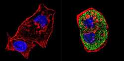
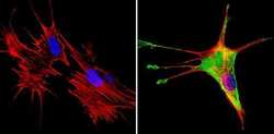
- Immunofluorescent analysis of SREBP1 (green) showing staining in the cytoplasm of C2C12 cells (right) compared to a negative control without primary antibody (left). Formalin-fixed cells were permeabilized with 0.1% Triton X-100 in TBS for 5-10 minutes and blocked with 3% BSA-PBS for 30 minutes at room temperature. Cells were probed with a SREBP1 polyclonal antibody (Product # PA1-337) in 3% BSA-PBS at a dilution of 1:100 and incubated overnight at 4ºC in a humidified chamber. Cells were washed with PBST and incubated with a DyLight-conjugated secondary antibody in PBS at room temperature in the dark. F-actin (red) was stained with a fluorescent red phalloidin and nuclei (blue) were stained with Hoechst or DAPI. Images were taken at a magnification of 60x.

- Submitted by
- Invitrogen Antibodies (provider)
- Main image
- Experimental details
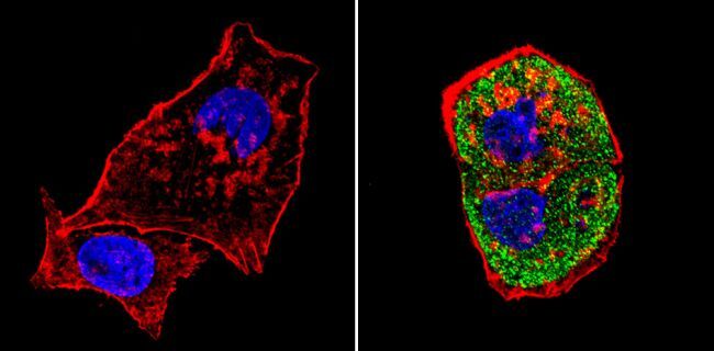
- Immunofluorescent analysis of SREBP1 (green) showing staining in the cytoplasm of HepG2 cells (right) compared to a negative control without primary antibody (left). Formalin-fixed cells were permeabilized with 0.1% Triton X-100 in TBS for 5-10 minutes and blocked with 3% BSA-PBS for 30 minutes at room temperature. Cells were probed with a SREBP1 polyclonal antibody (Product # PA1-337) in 3% BSA-PBS at a dilution of 1:100 and incubated overnight at 4ºC in a humidified chamber. Cells were washed with PBST and incubated with a DyLight-conjugated secondary antibody in PBS at room temperature in the dark. F-actin (red) was stained with a fluorescent red phalloidin and nuclei (blue) were stained with Hoechst or DAPI. Images were taken at a magnification of 60x.

- Submitted by
- Invitrogen Antibodies (provider)
- Main image
- Experimental details
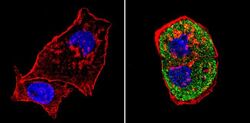
- Immunofluorescent analysis of SREBP1 (green) showing staining in the cytoplasm of NIH-3T3 cells (right) compared to a negative control without primary antibody (left). Formalin-fixed cells were permeabilized with 0.1% Triton X-100 in TBS for 5-10 minutes and blocked with 3% BSA-PBS for 30 minutes at room temperature. Cells were probed with a SREBP1 polyclonal antibody (Product # PA1-337) in 3% BSA-PBS at a dilution of 1:100 and incubated overnight at 4ºC in a humidified chamber. Cells were washed with PBST and incubated with a DyLight-conjugated secondary antibody in PBS at room temperature in the dark. F-actin (red) was stained with a fluorescent red phalloidin and nuclei (blue) were stained with Hoechst or DAPI. Images were taken at a magnification of 60x.

- Submitted by
- Invitrogen Antibodies (provider)
- Main image
- Experimental details
- Immunofluorescent analysis of SREBP1 (green) showing staining in the cytoplasm of HepG2 cells (right) compared to a negative control without primary antibody (left). Formalin-fixed cells were permeabilized with 0.1% Triton X-100 in TBS for 5-10 minutes and blocked with 3% BSA-PBS for 30 minutes at room temperature. Cells were probed with a SREBP1 polyclonal antibody (Product # PA1-337) in 3% BSA-PBS at a dilution of 1:100 and incubated overnight at 4ºC in a humidified chamber. Cells were washed with PBST and incubated with a DyLight-conjugated secondary antibody in PBS at room temperature in the dark. F-actin (red) was stained with a fluorescent red phalloidin and nuclei (blue) were stained with Hoechst or DAPI. Images were taken at a magnification of 60x.
- Submitted by
- Invitrogen Antibodies (provider)
- Main image

- Experimental details
- Immunofluorescent analysis of SREBP1 (green) showing staining in the cytoplasm of NIH-3T3 cells (right) compared to a negative control without primary antibody (left). Formalin-fixed cells were permeabilized with 0.1% Triton X-100 in TBS for 5-10 minutes and blocked with 3% BSA-PBS for 30 minutes at room temperature. Cells were probed with a SREBP1 polyclonal antibody (Product # PA1-337) in 3% BSA-PBS at a dilution of 1:100 and incubated overnight at 4ºC in a humidified chamber. Cells were washed with PBST and incubated with a DyLight-conjugated secondary antibody in PBS at room temperature in the dark. F-actin (red) was stained with a fluorescent red phalloidin and nuclei (blue) were stained with Hoechst or DAPI. Images were taken at a magnification of 60x.

- Submitted by
- Invitrogen Antibodies (provider)
- Main image
- Experimental details
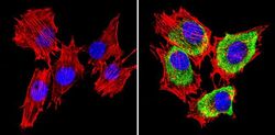
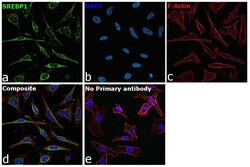
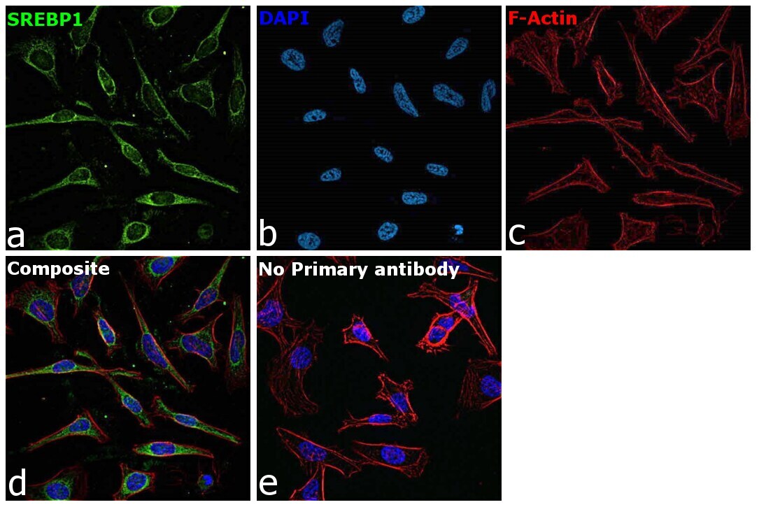
- Immunofluorescence analysis of SREBF1 was performed using 70% confluent log phase HeLa cells. The cells were fixed with 4% paraformaldehyde for 10 minutes, permeabilized with 0.1% Triton™ X-100 for 15 minutes, and blocked with 2% BSA for 1 hour at room temperature. The cells were labeled with SREBP1 Polyclonal Antibody (Product # PA1-337, 1:200) in 0.1% BSA, incubated at 4 degree celsius overnight and then labeled with Donkey anti-Rabbit IgG (H+L) Highly Cross-Adsorbed Secondary Antibody, Alexa Fluor™ Plus 488 (Product # A32790, 1:2,000), for 45 minutes at room temperature (Panel a: Green). Nuclei (Panel b:Blue) were stained with ProLong™ Diamond Antifade Mountant with DAPI (Product # P36962). F-actin (Panel c: Red) was stained with Rhodamine Phalloidin (Product # R415, 1:300). Panel d represents the merged image showing predominant cytoplasmic localization. Panel e represents control cells with no primary antibody to assess background. The images were captured at 40X magnification.
Supportive validation
- Submitted by
- Invitrogen Antibodies (provider)
- Main image

- Experimental details
- NULL
- Submitted by
- Invitrogen Antibodies (provider)
- Main image

- Experimental details
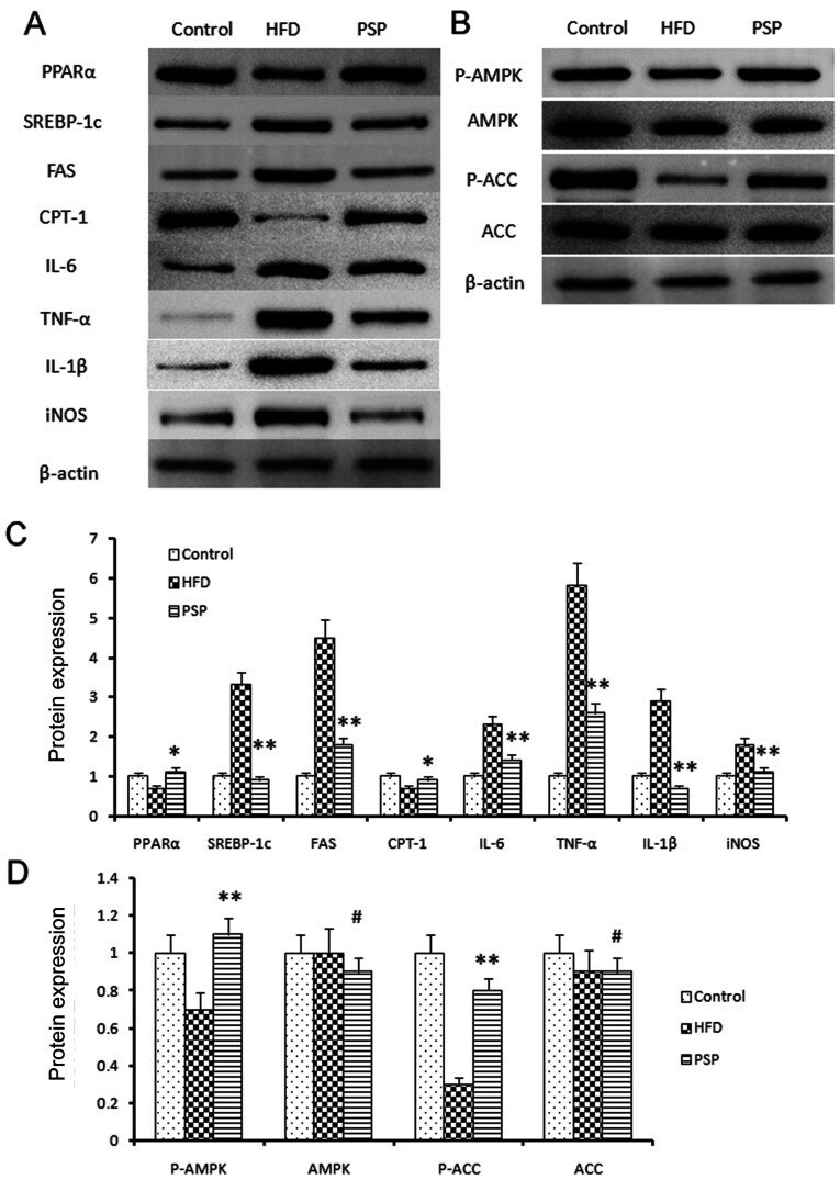
- Figure 5. Signal pathway changes and lipid-associated genes and inflammatory cytokine protein changes. (A) Western blotting was performed to reveal the protein expressions of the lipid and inflammatory genes as well as the key genes of the AMPK signal pathway. Lipid synthesis-associated genes and inflammatory genes were inhibited by PSP. (B) P-AMPK and the downstream gene p-ACC was clearly activated by PSP, which demonstrated that the PSP could ameliorate HFD-induced mouse obesity through activating the AMPK signal pathway. (C) Semi-quantification of protein expression levels from part (A) (D) Semi-quantification of protein expression levels from part (B) # P>0.05, *P
- Submitted by
- Invitrogen Antibodies (provider)
- Main image
- Experimental details
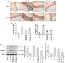
- Figure 5. Effects of GA on macrophage activation, alpha-SMA expression, FAS and SREBP-1c expression in DM-AS rats. (A) Immunohistochemistry was used to detect CD68 and alpha-SMA expression in aortic tissues (magnification, x200). (B) Quantitative analysis of CD68 and alpha-SMA expression from (A). (C) Western blotting detected the expression of FAS and SREBP-1c. ***P

- Submitted by
- Invitrogen Antibodies (provider)
- Main image
- Experimental details
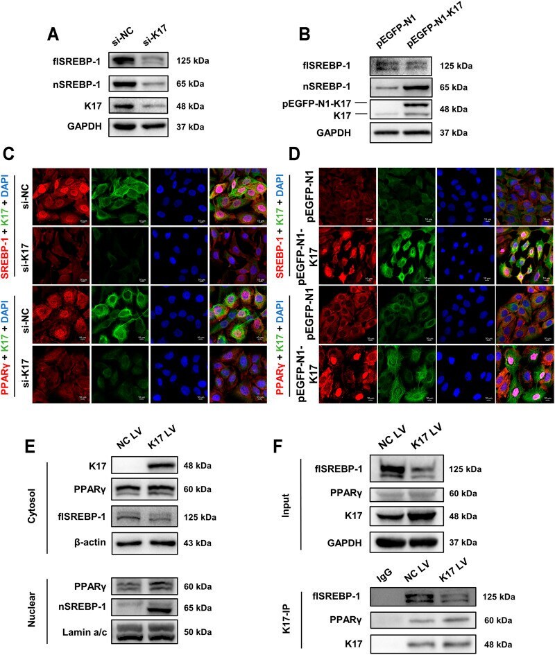
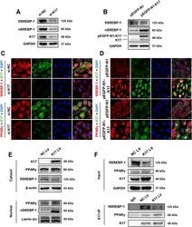
- FIGURE 5 K17 alters the subcellular localization of SREBP-1 and PPARgamma. (A,B) The protein levels of full-length SPEBP-1 (flSREBP-1) and nuclear SPEBP-1 (nSREBP-1) in HaCaT cells were measured after transfection with K17 siRNA or pEGFP-N1-K17. (C,D) Immunofluorescence analysis of FASN and PPARgamma subcellular localization in HaCaT cells after transfection with K17 siRNA or pEGFP-N1-K17. Scale bars = 10 mum. (E) HaCaT cells were treated with pCMV6-XL5-K17 (K17 LV) and a nuclear fraction was prepared. SREBP-1 and PPARgamma protein was quantitated by Western blot. Lamin A/C and beta-actin were used as loading controls for nuclear and cytosol fractions, respectively. (F) Cells were co-immunoprecipitated with anti-K17 antibody and SREBP-1 and PPARgamma proteins were detected by Western blot. Data are expressed as means +- standard error of the mean from three independent experiments. * p < 0.05.