 Explore
 Validate
 Learn
Learn Western blot
Antibody data
- Antibody Data
- Antigen structure
- References [2]
- Comments [0]
- Validations
- Western blot [2]
Submit
Validation data
Reference
Comment
Report error
- Product number
- MAB6434 - Provider product page

- Provider
- Novus Biologicals
- Product name
- Rat Monoclonal VCAM-1/CD106 Antibody
- Antibody type
- Monoclonal
- Description
- Protein A or G purified from hybridoma culture supernatant. Detects mouse VCAM-1 in direct ELISAs and Western blots. Does not cross-react with recombinant human (rh) VCAM-1, rmICAM-1, rhICAM-2, rmICAM-2, rhICAM-3, or rhPECAM.
- Reactivity
- Mouse
- Host
- Rat
- Conjugate
- Unconjugated
- Isotype
- IgG
- Vial size
- 500 ug
- Storage
- Use a manual defrost freezer and avoid repeated freeze-thaw cycles. 12 months from date of receipt, -20 to -70 degreesC as supplied. 1 month, 2 to 8 degreesC under sterile conditions after reconstitution. 6 months, -20 to -70 degreesC under sterile conditions after reconstitution.
Submitted references Lipopolysaccharides, but not Angiotensin ll, lnduces Direct Pro-lnflammatory Effects in Cultured Mouse Arteries and Human Endothelial and Vascular Smooth Muscle Cells.
Noninvasive ultrasound molecular imaging of the effect of statins on endothelial inflammatory phenotype in early atherosclerosis.
Outzen EM, Zaki M, Mehryar R, Abdolalizadeh B, Sajid W, Boonen HC, Sams A, Sheykhzade M
Basic & clinical pharmacology & toxicology 2017 Apr;120(4):335-347
Basic & clinical pharmacology & toxicology 2017 Apr;120(4):335-347
Noninvasive ultrasound molecular imaging of the effect of statins on endothelial inflammatory phenotype in early atherosclerosis.
Khanicheh E, Mitterhuber M, Xu L, Haeuselmann SP, Kuster GM, Kaufmann BA
PloS one 2013;8(3):e58761
PloS one 2013;8(3):e58761
No comments: Submit comment
Supportive validation
- Submitted by
- Novus Biologicals (provider)
- Main image
- Experimental details
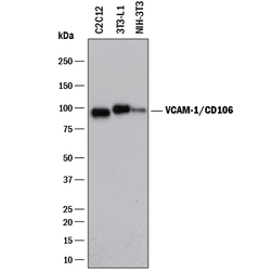
- Detection of Mouse VCAM-1/CD106 by Western Blot. Western blot shows lysates of 3T3-L1 mouse embryonic fibroblast adipose-like cell line, C2C12 mouse myoblast cell line, and NIH-3T3 mouse embryonic fibroblast cell line. PVDF membrane was probed with 2 µg/mL of Rat Anti-Mouse VCAM-1/CD106 Monoclonal Antibody (Catalog # MAB6434) followed by HRP-conjugated Anti-Rat IgG Secondary Antibody (Catalog # HAF005). A specific band was detected for VCAM-1/ CD106 at approximately 95 kDa (as indicated). This experiment was conducted under reducing conditions and using Immunoblot Buffer Group 1.

- Submitted by
- Novus Biologicals (provider)
- Main image
- Experimental details
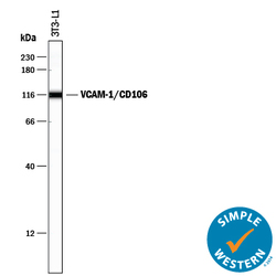
- Detection of Mouse VCAM-1/CD106 by Simple WesternTM. Simple Western lane view shows lysate of 3T3-L1 mouse embryonic fibroblast adipose-like cell line, loaded at 0.2 mg/mL. A specific band was detected for VCAM-1/CD106 at approximately 117 kDa (as indicated) using 100 µg/mL of Rat Anti-Mouse VCAM-1/CD106 Monoclonal Antibody (Catalog # MAB6434) followed by 1:50 dilution of HRP-conjugated Anti-Rat IgG Secondary Antibody (Catalog # HAF005). This experiment was conducted under reducing conditions and using the 12-230 kDa separation system.
