 Explore
 Validate
 Learn
Learn Flow cytometry
Antibody data
- Antibody Data
- Antigen structure
- References [9]
- Comments [0]
- Validations
- Flow cytometry [2]
- Other assay [11]
Submit
Validation data
Reference
Comment
Report error
- Product number
- 13-1069-82 - Provider product page

- Provider
- Invitrogen Antibodies
- Product name
- CD106 (VCAM-1) Monoclonal Antibody (STA), Biotin, eBioscience™
- Antibody type
- Monoclonal
- Antigen
- Other
- Description
- Description: The STA monoclonal antibody reacts with human CD106 (Vascular Cell Adhesion Molecule-1, VCAM-1), a 110 kDa transmembrane glycoprotein expressed by myeloid lineage and bone marrow stromal cells. Endothelial cells constitutively express low levels of CD106 and upregulate it upon cytokine stimulation. CD106 binds to Integrin alpha4beta1 (VLA-4, CD49d/CD29) and Integrin alpha4beta7 (LPAM-1). Cytokine-mediated upregulation of CD106 on endothelial cells suggests a role for this antigen in the inflammatory response. Applications Reported: STA has been reported for use in flow cytometric analysis. Applications Tested: The STA antibody has been tested by flow cytometric analysis of TNF alpha-activated HUVEC cells. This can be used at less than or equal to 0.5 µg per test. A test is defined as the amount (µg) of antibody that will stain a cell sample in a final volume of 100 µL. Cell number should be determined empirically but can range from 10^5 to 10^8 cells/test. It is recommended that the antibody be carefully titrated for optimal performance in the assay of interest. Filtration: 0.2 µm post-manufacturing filtered.
- Reactivity
- Human
- Host
- Mouse
- Conjugate
- Biotin
- Isotype
- IgG
- Antibody clone number
- STA
- Vial size
- 100 µg
- Concentration
- 0.5 mg/mL
- Storage
- 4°C, store in dark, DO NOT FREEZE!
Submitted references Multimodal CRISPR perturbations of GWAS loci associated with coronary artery disease in vascular endothelial cells.
Colonization of dermal arterioles by Neisseria meningitidis provides a safe haven from neutrophils.
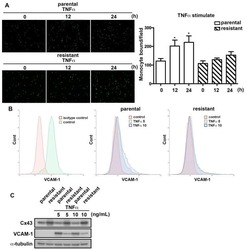
Differential Characterization of Temozolomide-Resistant Human Glioma Cells.
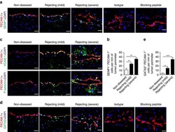
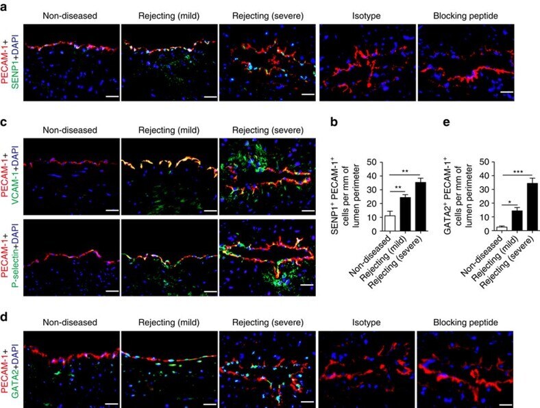
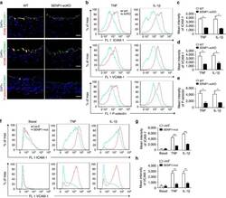
The critical role of SENP1-mediated GATA2 deSUMOylation in promoting endothelial activation in graft arteriosclerosis.
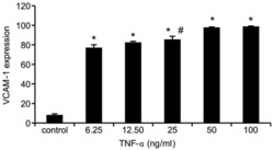
TNF-α enhances vascular cell adhesion molecule-1 expression in human bone marrow mesenchymal stem cells via the NF-κB, ERK and JNK signaling pathways.
Effect of nicotine and porphyromonas gingivalis lipopolysaccharide on endothelial cells in vitro.
Cytokine induction of VCAM-1 but not IL13Rα2 on glioma cells: a tale of two antibodies.
The CD14+CD16+ inflammatory monocyte subset displays increased mitochondrial activity and effector function during acute Plasmodium vivax malaria.
Effects of Aggregatibacter actinomycetemcomitans leukotoxin on endothelial cells.
Wünnemann F, Fotsing Tadjo T, Beaudoin M, Lalonde S, Lo KS, Kleinstiver BP, Lettre G
PLoS genetics 2023 Mar;19(3):e1010680
PLoS genetics 2023 Mar;19(3):e1010680
Colonization of dermal arterioles by Neisseria meningitidis provides a safe haven from neutrophils.
Manriquez V, Nivoit P, Urbina T, Echenique-Rivera H, Melican K, Fernandez-Gerlinger MP, Flamant P, Schmitt T, Bruneval P, Obino D, Duménil G
Nature communications 2021 Jul 27;12(1):4547
Nature communications 2021 Jul 27;12(1):4547
Differential Characterization of Temozolomide-Resistant Human Glioma Cells.
Lai SW, Huang BR, Liu YS, Lin HY, Chen CC, Tsai CF, Lu DY, Lin C
International journal of molecular sciences 2018 Jan 2;19(1)
International journal of molecular sciences 2018 Jan 2;19(1)
The critical role of SENP1-mediated GATA2 deSUMOylation in promoting endothelial activation in graft arteriosclerosis.
Qiu C, Wang Y, Zhao H, Qin L, Shi Y, Zhu X, Song L, Zhou X, Chen J, Zhou H, Zhang H, Tellides G, Min W, Yu L
Nature communications 2017 Jun 1;8:15426
Nature communications 2017 Jun 1;8:15426
TNF-α enhances vascular cell adhesion molecule-1 expression in human bone marrow mesenchymal stem cells via the NF-κB, ERK and JNK signaling pathways.
Lu ZY, Chen WC, Li YH, Li L, Zhang H, Pang Y, Xiao ZF, Xiao HW, Xiao Y
Molecular medicine reports 2016 Jul;14(1):643-8
Molecular medicine reports 2016 Jul;14(1):643-8
Effect of nicotine and porphyromonas gingivalis lipopolysaccharide on endothelial cells in vitro.
An N, Andrukhov O, Tang Y, Falkensammer F, Bantleon HP, Ouyang X, Rausch-Fan X
PloS one 2014;9(5):e96942
PloS one 2014;9(5):e96942
Cytokine induction of VCAM-1 but not IL13Rα2 on glioma cells: a tale of two antibodies.
Mahadev V, Starr R, Wright SL, Martinez C, Jensen MC, Barish ME, Forman SJ, Brown CE
PloS one 2014;9(5):e95123
PloS one 2014;9(5):e95123
The CD14+CD16+ inflammatory monocyte subset displays increased mitochondrial activity and effector function during acute Plasmodium vivax malaria.
Antonelli LR, Leoratti FM, Costa PA, Rocha BC, Diniz SQ, Tada MS, Pereira DB, Teixeira-Carvalho A, Golenbock DT, Gonçalves R, Gazzinelli RT
PLoS pathogens 2014 Sep;10(9):e1004393
PLoS pathogens 2014 Sep;10(9):e1004393
Effects of Aggregatibacter actinomycetemcomitans leukotoxin on endothelial cells.
Dietmann A, Millonig A, Combes V, Couraud PO, Kachlany SC, Grau GE
Microbial pathogenesis 2013 Aug-Sep;61-62:43-50
Microbial pathogenesis 2013 Aug-Sep;61-62:43-50
No comments: Submit comment
Supportive validation
- Submitted by
- Invitrogen Antibodies (provider)
- Main image

- Experimental details
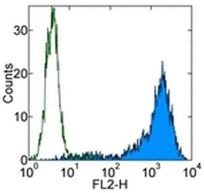
- Staining of TNF alpha-stimulated Human Umbilical Vein Endothelial Cells (HUVEC) cells with 0.5 µg of Mouse IgG1 kappa Isotype Control Biotin (Product # 13-4714-85) (open histogram) or 0.5 µg of Anti-Human CD106 (VCAM-1) Biotin (filled histogram) followed by Streptavidin PE (Product # 12-4317-87). Total viable cells were used for analysis.
- Conjugate
- Biotin
- Submitted by
- Invitrogen Antibodies (provider)
- Main image

- Experimental details
- Staining of TNF alpha-stimulated Human Umbilical Vein Endothelial Cells (HUVEC) cells with 0.5 µg of Mouse IgG1 kappa Isotype Control Biotin (Product # 13-4714-85) (open histogram) or 0.5 µg of Anti-Human CD106 (VCAM-1) Biotin (filled histogram) followed by Streptavidin PE (Product # 12-4317-87). Total viable cells were used for analysis.

Supportive validation
- Submitted by
- Invitrogen Antibodies (provider)
- Main image
- Experimental details
- NULL
- Conjugate
- Biotin

- Submitted by
- Invitrogen Antibodies (provider)
- Main image
- Experimental details
- NULL
- Conjugate
- Biotin
- Submitted by
- Invitrogen Antibodies (provider)
- Main image
- Experimental details
- NULL
- Conjugate
- Biotin

- Submitted by
- Invitrogen Antibodies (provider)
- Main image
- Experimental details
- Figure 1 Enhanced expression of endothelial SENP1 and GATA2 correlates with graft arteriosclerosis (GA) progression. Similarly sized human coronary arteries with GA from chronically rejecting heart allografts or without disease from non-transplanted hearts were collected and evaluated by histological analysis. ( a , b ) Dramatically increased expression of endothelial SENP1 was detected in the diseased vessel wall. Endothelial SENP1 expression is demonstrated by immunofluorescence analysis of coronary artery cross-sections that were stained for SENP1 and the endothelial marker PECAM-1 with DAPI labelling of the nuclei. Representative images are shown in ( a ) with quantification data in ( b ). Bar represents 50 mum. ( c ) Induction of endothelial adhesion molecules resulted in a similar augmented pattern as endothelial SENP1. Representative images of immunofluorescence staining for VCAM-1 or P-selectin and PECAM-1 in coronary arteries with DAPI counterstaining are shown. Bar represents 50 mum. ( d , e ) Expression of endothelial GATA2 was elevated with the progression of aggravated rejection. Representative images of the immunofluorescence staining of coronary arteries for GATA2 and PECAM-1 together with DAPI nuclear staining are shown in ( d ) with quantification data in ( e ). Negative SENP1 and GATA2 staining in the isotype or blocking peptide controls are also shown in ( a ) and ( d ). Bar represents 50 mum. Data presented in ( b , e ) are the mean+-s.e.m. from five separ
- Conjugate
- Biotin
- Submitted by
- Invitrogen Antibodies (provider)
- Main image
- Experimental details
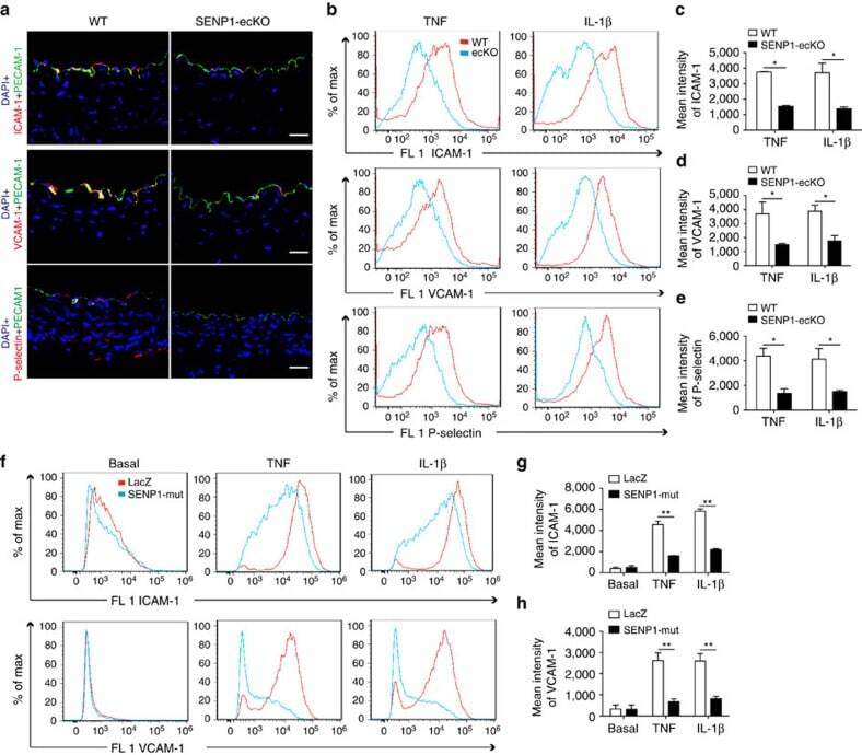
- Figure 3 Loss of endothelial SENP1 inhibits EC activation. ( a ) Grafts from WT or SENP1-ecKO mice were harvested 3 days post-transplantation. The induction of endothelial adhesion molecules was demonstrated by immunofluorescence staining of ICAM-1, VCAM-1, or P-selectin and PECAM-1 with DAPI labelling of the nuclei. Bar represents 50 mum. ( b - e ) Attenuated induction of adhesion molecules in SENP1-ecKO MAECs. Flow cytometry analysis of ICAM-1, VCAM-1 and P-selectin in MAECs isolated from WT or SENP1-ecKO mice after TNF or IL-1beta treatment. Representative histograms are shown in ( b ) with the quantification of mean intensity in ( c - e ). ( f - h ) Overexpression of the catalytically inactive form of SENP1 (SENP1-Mut) inhibits the induction of adhesion molecules in HUVECs. HUVECs were infected by Ad-SENP1-Mut or vector control (Ad-LacZ) for 24 h, treated with pro-inflammatory cytokines and analysed by flow cytometry in the same way as MAECs. Representative histograms of ICAM-1 and VCAM-1 are shown in ( f ) with the quantification of mean intensity in ( g , h ). Data are presented as the mean+-s.e.m. from at least three independent experiments. * P
- Conjugate
- Biotin
- Submitted by
- Invitrogen Antibodies (provider)
- Main image
- Experimental details
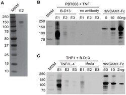
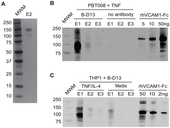
- Figure 4 The B-D13 antibody recognizes VCAM-1, not IL13Ralpha2. (A) Silver stain gel of the second elution (E2) from the B-D13 immunoprecipitation of PBT008 stimulated overnight with TNF. (B) Western blot detecting B-D13 pull-down of VCAM-1 in immunoprecipitation eluates (E1, E2, E3) for PBT008 cells stimulated overnight with TNF. VCAM-1 was not immunoprecipitated in the absence of B-D13 antibody (no antibody: E1, E2, E3). Titrated soluble recombinant human VCAM-1-Fc shows specificity of the VCAM-1 antibody. (C) Western blot detecting B-D13 pull-down of VCAM-1 in immunoprecipitation eluates (E1, E2, E3) for THP-1 cells in both media and cytokine overnight-stimulated conditions. All data are representative of 2 separate experiments.
- Conjugate
- Biotin
- Submitted by
- Invitrogen Antibodies (provider)
- Main image
- Experimental details
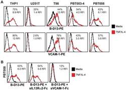
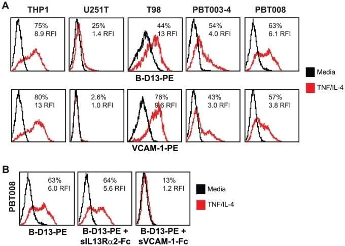
- Figure 5 Antibody immunoreactivity, and soluble ligand competition confirms B-D13-PE specificity for VCAM-1. (A) Flow cytometric analysis of monocytic line THP-1 and various glioma lines with B-D13-PE (top), or PE-conjugated anti-VCAM-1 (VCAM-1-PE, bottom), after overnight culture in media alone (black histogram) versus cytokine (TNF/IL-4; red histogram) conditions. Data are representative of at least 2 separate experiments. (B) PBT008 cells cultured overnight in media alone (black histogram) versus cytokine (TNF/IL-4; red histogram) conditions were stained with B-D13-PE antibody that was pre-incubated with soluble recombinant human IL13Ralpha-Fc or VCAM-1-Fc. As a control, cells were stained with B-D13-PE alone. Data are representative of 2 separate experiments.
- Conjugate
- Biotin
- Submitted by
- Invitrogen Antibodies (provider)
- Main image
- Experimental details
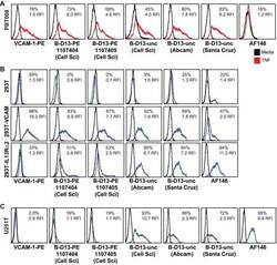
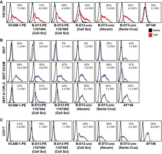
- Figure 6 Evaluation of additional commercially available B-D13 antibodies. (A) PBT008 cells that had been cultured overnight in media alone (black histogram) versus cytokine (TNF; red histogram) conditions, (B) Parental 293T cells or 293T cells engineered to express either VCAM-1 or IL13Ralpha2, and (C) U251T cells were stained with VCAM-1-PE, AF146 or various B-D13 reagents - two lots of PE-conjugated B-D13 antibody (B-D13-PE; Cell Sciences) and two to three unconjugated B-D13 antibodies (B-D13-unc) purchased from either Cell Sciences (Cell Sci), Abcam or Santa Cruz as indicated. (B, C) Black histograms represent staining with istoype control antibody or SA-PE alone.
- Conjugate
- Biotin
- Submitted by
- Invitrogen Antibodies (provider)
- Main image
- Experimental details
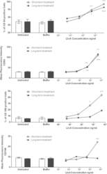
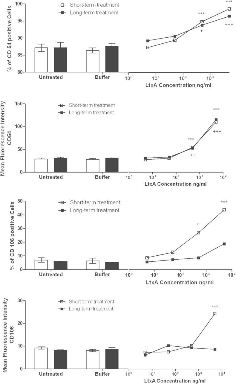
- Fig. 7 Activation of adhesion molecules. hCMEC/D3 cells were seeded in the presence of a single LtxA dose or buffer (long-term) or confluent hCMEC/D3 monolayers were treated with LtxA or buffer for 16 h (short-term). When untreated cells reached confluency (long-term), or after overnight stimulation (short-term), cells were harvested and stained for cellular adhesion molecules CD54 (ICAM-1) or CD106 (VCAM-1). Percentages of CD54 or CD106 positive cells as well as CD54 and CD106 mean fluorescence intensity are shown for untreated, buffer-treated, or LtxA-treated cells. Data represents means and SEM. ANOVA was used for each treatment group. * refers to the short-term treatment groups, + refers to the long-term treatment groups. Figure shows significances for testing against buffer-treated group only. */+ p < .05, **/++ p < .01, ***/+++ p < .001.
- Conjugate
- Biotin
- Submitted by
- Invitrogen Antibodies (provider)
- Main image
- Experimental details
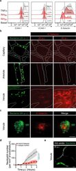
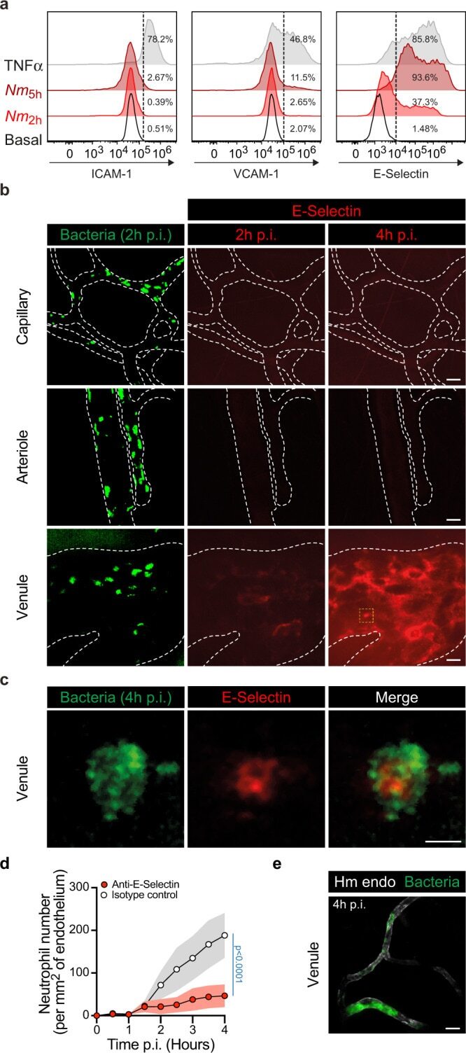
- Fig. 7 E-selectin endothelium surface expression is differentially upregulated according to the vascular bed upon infection. a Flow cytometry analysis of cell surface expression of ICAM-1, VCAM-1, and E-selectin (CD62E) on HUVEC cells under resting conditions (Basal, open histograms) or following infection with Neisseria meningitidis for 2 h ( Nm 2h , red histograms) or 5 h ( Nm 5h , dark-red histograms) or an overnight incubation with 20 ng ml -1 TNFalpha gray histograms). Data are representatives of N = 3 independent experiments. The percentages of positive cells (above the dashed lines) are shown per condition and marker. b In vivo expression of E-selectin at the surface of different human vessel types (capillary, arteriole, and venule) following two (2 h p.i.) and four (4 h p.i.) hours of infection. Images (maximum intensity z-projection) are representative of N = 3 infected mice imaged independently. GFP-expressing Neisseria meningitidis appears in green and E-selectin in red following in vivo labeling by i.v. injection of PE-labeled anti-CD62E monoclonal antibody. Dashed lines delineate human vessels (UEA-1 lectin). Scale bar, 20 um. c High-magnification view (yellow dashed square in panel b ) of a bacterial microcolony at 4 h p.i. on the venular endothelium surface and the local upregulation of E-selectin expression. Scale bar, 5 um. d Movies obtained from intravital imaging were used to quantify the numbers of neutrophils per square millimeter of venular endothelium d
- Conjugate
- Biotin
- Submitted by
- Invitrogen Antibodies (provider)
- Main image
- Experimental details
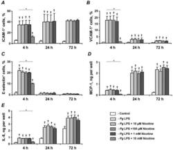
- Figure 5 Effect of nicotine on the P. gingivalis LPS-induced protein expression of pro-inflammatory mediators in HUVECs. HUVECs were stimulated by P. gingivalis LPS in the presence or absence of nicotine (10 uM-10 mM) for 4, 24, and 72 h. After stimulation, the surface expression levels of ICAM-1 (A), VCAM-1 (B), and E-selectin (C) were measured by flow cytometry, and the quantity of MCP-1 (D) and IL-8 (E) in conditioned media was measured by ELISA. Each value represents mean +-SD of three independent assays. Non-stimulated HUVECs were used as a control. The protein expression levels of pro-inflammatory mediators were not analyzed after stimulation with 10-mM nicotine for 24 and 72 h because the cells were not viable. * - significantly different between groups, p
- Conjugate
- Biotin
