Explore
 Validate
 Learn
Learn Western blot
 ELISA
ELISAAntibody data
- Antibody Data
- Antigen structure
- References [2]
- Comments [0]
- Validations
- Western blot [4]
- Immunocytochemistry [1]
- Other assay [2]
Submit
Validation data
Reference
Comment
Report error
- Product number
- M808 - Provider product page

- Provider
- Invitrogen Antibodies
- Product name
- VEGF Monoclonal Antibody (16F1)
- Antibody type
- Monoclonal
- Antigen
- Recombinant full-length protein
- Description
- The VEGF-A monoclonal antibody clone # 16F1 (Product # M808) has successfully been paired as the coating antibody in a sandwich ELISA with detection antibody P802B (biotinylated conjugate of Product # P802). Typical dilutions for sandwich ELISA include 1 µg/mL for coating and 0.125-0.5 µg/mL for detection.
- Reactivity
- Human
- Host
- Mouse
- Isotype
- IgG
- Antibody clone number
- 16F1
- Vial size
- 200 µg
- Concentration
- 1 mg/mL
- Storage
- -20°C
Submitted references Qi-Long-Tian formula extract alleviates symptoms of acute high-altitude diseases via suppressing the inflammation responses in rat.
Upregulation of OTUD7B (Cezanne) Promotes Tumor Progression via AKT/VEGF Pathway in Lung Squamous Carcinoma and Adenocarcinoma.
Fu X, Yang C, Chen B, Zeng K, Chen S, Fu Y
Respiratory research 2021 Feb 12;22(1):52
Respiratory research 2021 Feb 12;22(1):52
Upregulation of OTUD7B (Cezanne) Promotes Tumor Progression via AKT/VEGF Pathway in Lung Squamous Carcinoma and Adenocarcinoma.
Lin DD, Shen Y, Qiao S, Liu WW, Zheng L, Wang YN, Cui N, Wang YF, Zhao S, Shi JH
Frontiers in oncology 2019;9:862
Frontiers in oncology 2019;9:862
No comments: Submit comment
Supportive validation
- Submitted by
- Invitrogen Antibodies (provider)
- Main image

- Experimental details
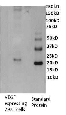
- Western blot analysis of Human VEGF was performed by loading 0. 5 µg of VEGF transfected 293T cell culture supernatant and recombinant human VEGF onto a 4-12% Tris-HCl polyacrylamide gel. Proteins were transferred to a PVDF membrane and blocked with 3% BSA/TBST for at least 1 hour. The membrane was probed with a human VEGF monoclonal antibody (Product # M808) at a dilution of 1 µg/mL overnight at 4°C on a rocking platform, washed in TBS-0.1%Tween 20 and probed with a goat anti- mouse-HRP secondary antibody (Product # 31430) at a dilution of 1:15,000 for at least one hour. Chemiluminescent detection was performed using SuperSignal West Dura (Product # 34075).
- Submitted by
- Invitrogen Antibodies (provider)
- Main image

- Experimental details
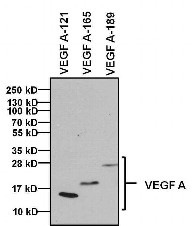
- Western blot analysis of Vascular Endothelial Growth Factor (VEGF) was performed by loading 1 µg of indicated recombinant VEGF isoforms, and 10 µL of PageRuler PlusPrestained Protein Ladder (Product # 26619) per well onto a 4-20% Tris-Glycine polyacrylamide gel. Proteins were transferred to a PVDF membrane (Product # 88518) using the G2 Fast Blotter (Product # 62288) and blocked with 5% Milk/TBST for at least 1 hour at room temperature. VEGF was detected at 14 kD, 19 kD and 22-30 kD using a VEGF monoclonal antibody (Product # M808) at a dilution of 1:1000 in blocking buffer overnight at 4C on a rocking platform, followed by an HRP-conjugated goat anti-mouse IgG (Fc) secondary antibody (Product # 31437) at a dilution of 1:20,000 for at least 1 hour. Chemiluminescent detection was performed using SuperSignal West Dura (Product # 34076).
- Submitted by
- Invitrogen Antibodies (provider)
- Main image

- Experimental details
- Western blot analysis of Vascular Endothelial Growth Factor (VEGF) was performed by loading 1 µg of indicated recombinant VEGF isoforms, and 10 µL of PageRuler PlusPrestained Protein Ladder (Product # 26619) per well onto a 4-20% Tris-Glycine polyacrylamide gel. Proteins were transferred to a PVDF membrane (Product # 88518) using the G2 Fast Blotter (Product # 62288) and blocked with 5% Milk/TBST for at least 1 hour at room temperature. VEGF was detected at 14 kD, 19 kD and 22-30 kD using a VEGF monoclonal antibody (Product # M808) at a dilution of 1:1000 in blocking buffer overnight at 4C on a rocking platform, followed by an HRP-conjugated goat anti-mouse IgG (Fc) secondary antibody (Product # 31437) at a dilution of 1:20,000 for at least 1 hour. Chemiluminescent detection was performed using SuperSignal West Dura (Product # 34076).

- Submitted by
- Invitrogen Antibodies (provider)
- Main image

- Experimental details
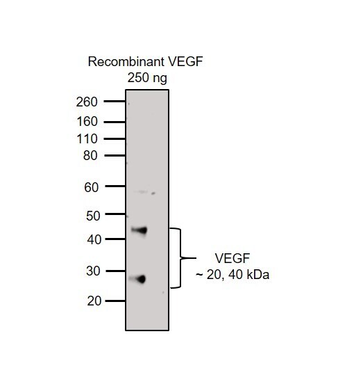
- Western blot was performed using Anti-VEGF Monoclonal Antibody (16F1) (Product # M808) and 20 and 40 kDa bands corresponding to VEGF were observed. Recombinant VEGF protein (250 ng) was electrophoresed using NuPAGE™ 4-12% Bis-Tris Protein Gel (Product # NP0322BOX). Resolved proteins were then transferred onto a Nitrocellulose membrane (Product # IB23002) by iBlot® 2 Dry Blotting System (Product # IB21001). The blot was probed with the primary antibody (1 µg/ml) and detected by chemiluminescence with Goat anti-Mouse IgG (H+L) Superclonal™ Recombinant Secondary Antibody, HRP (Product # A28177, 1:4000) using the iBright FL 1000 (Product # A32752). Chemiluminescent detection was performed using Novex® ECL Reagent Kit (Product # WP20005).
Supportive validation
- Submitted by
- Invitrogen Antibodies (provider)
- Main image
- Experimental details
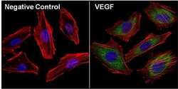
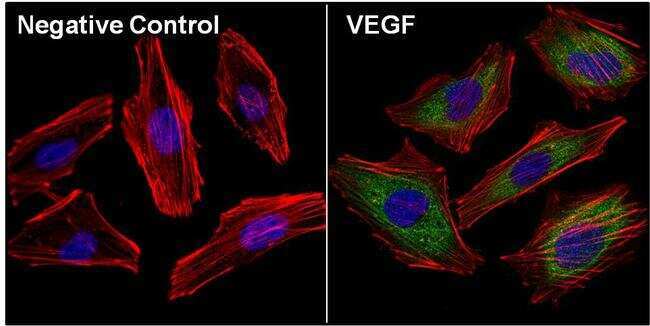
- Immunofluorescent analysis of Vascular Endothelial Growth Factor (VEGF, green) in Hela cells. The cells were fixed with 4% paraformaldehyde, permeabilized with 0.1% Triton X-100 in PBS, and blocked with 3% Blocker BSA (Product # 37525) for 30 minutes at room temperature. Cells were stained with (left panel) or without (right panel) a VEGF polyclonal antibody (Product # M808) at a dilution of 1:40 for at least 1 hour at room temperature, and then incubated with a Dylight 488 goat anti-mouse IgG secondary antibody at a dilution of 1:1000 for 45 minutes at room temperature. F-actin (both panels, red) was stained by Dylight 554 Phalloidin (Product # 21834) and nuclei (both panels, blue) were stained with DAPI (Product # 46190). Images were taken at 60X magnification.
Supportive validation
- Submitted by
- Invitrogen Antibodies (provider)
- Main image

- Experimental details
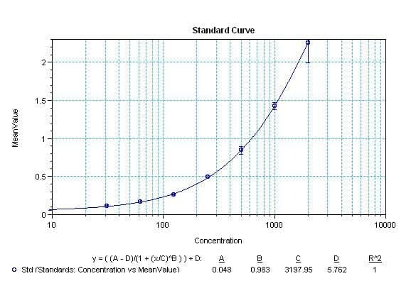
- Sandwich ELISA analysis of Human VEGF was performed using the Thermo Scientific ELISA Kit (Product # EHVEGF) by coating a blank 96-well microtiter plate with 100 µL per well of Human VEGF moncolonal antibody (Product # M808) in duplicate at 1, 3, 4, 5, 7, and 9 µg/mL in DPBS (Product # 28374) and incubating for 12-18 hours at 4°C. The plate was aspirated and blocked with 300 µL per well of 4% BSA and 5% sucrose in DPBS for 1 hour at room temperature. Human VEGF recombinant protein (Product # SVEGF) at 50 µL per well (in addition to 50 µL of sample diluent per well) was added in duplicate at 2000, 1000, 500, 250, 125, 62.5, 31.25, and 0pg/mL for 2 hours at room temperature. The plate was washed with ELISA Wash Buffer (Product # N503) and incubated with 100 µL of Human VEGF biotinylated polyclonal antibody (Product # P802B) in all applicable wells at 0.5 µg/mL for 1 hour at room temperature. The plate was washed and incubated with 100 µL per well of Streptavidin-HRP (Product # N504) in all test wells at 1:40,000 dilution for 30 minutes at room temperature and then washed and incubated with 100 µL per well of TMB substrate (Product # 34028) for 30 minutes at room temperature in the dark. The plate was stopped with 0.16M sulfuric acid (Product # N600). Absorbances were read on a spectrophotometer at 450-550 nm.
- Submitted by
- Invitrogen Antibodies (provider)
- Main image
- Experimental details
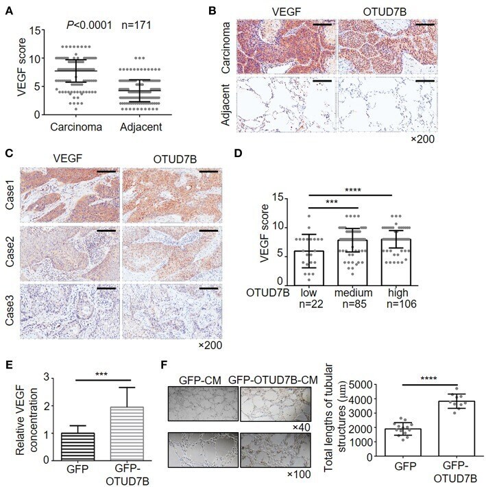
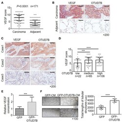
- Figure 4 Positive correlation of OTUD7B with VEGF expression and its role in VEGF production and angiogenesis. (A) The expression scores of VEGF ( n = 171) were compared between NSCLC tumors and matched adjacent normal tissue using paired t- test. Data are shown as mean +- s.d. (B) Representative images from IHC staining of VEGF in the same NSCLC tumor. Magnification, x200; Scale bars, 100 mum. (C) Representative images from IHC staining of VEGF and OTUD7B expression in two serial sections of the same tumor from three cases. Magnification, x200; Scale bars, 100 mum. (D) Scatter dot plots of VEGF expression in NSCLC from 213 subjects. The subjects were divided into three groups based on OTUD7B expression scores in the tumors, representing low, medium and high expression of OTUD7B. Data were analyzed by one-way ANOVA and Tukey's multiple comparisons test. (E) Media from GFP- or GFP-OTUD7B-overexpressing NCI-H358 cells was removed, washed in RPMI-1640 with 0.5% FBS and incubated for an additional 24 h in RPMI-1640 with 0.5% FBS. GFP-conditioned media (GFPCM) and GFP-OTUD7B-conditioned media (GFP-OTUD7B-CM) were collected, and VEGF expression were analyzed by ELISA. Absorbance was measured at 450 nm. (F) EA.hy926 endothelial cells were pretreated with GFP-CM and GFP-OTUD7B-CM for 24 h. Subsequently, pretreated EA.hy926 cells were seeded on Matrigel for 8 h to observe tube formation. Representative photographs are shown (left). Tube lengths were quantitated using IMAGE-PRO PLUS so