Explore
 Validate
 Learn
Learn Western blot
Antibody data
- Antibody Data
- Antigen structure
- References [0]
- Comments [0]
- Validations
- Western blot [3]
- Immunocytochemistry [2]
- Other assay [1]
Submit
Validation data
Reference
Comment
Report error
- Product number
- P802 - Provider product page

- Provider
- Invitrogen Antibodies
- Product name
- VEGF Polyclonal Antibody
- Antibody type
- Polyclonal
- Antigen
- Recombinant full-length protein
- Description
- The VEGF-A monoclonal antibody clone # 16F1 (Product # M808) has successfully been paired as the coating antibody in a sandwich ELISA with detection antibody P802B (biotinylated conjugate of Product # P802).
- Concentration
- 1.0 mg/mL
No comments: Submit comment
Supportive validation
- Submitted by
- Invitrogen Antibodies (provider)
- Main image
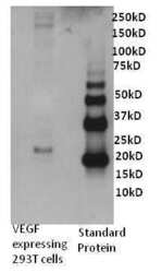
- Experimental details
- Western blot analysis of Human VEGF was performed by loading 0. 5 µg of VEGF transfected 293T cell culture supernatant and recombinant human VEGF onto a 4-12% Tris-HCl polyacrylamide gel. Proteins were transferred to a PVDF membrane and blocked with 3% BSA/TBST for at least 1 hour. The membrane was probed with a human VEGF polyclonal antibody (Product # P802) at a dilution of 1 µg/mL overnight at 4°C on a rocking platform, washed in TBS-0.1%Tween 20 and probed with a goat anti- rabbit-HRP secondary antibody (Product # 31460) at a dilution of 1:15,000 for at least one hour. Chemiluminescent detection was performed using SuperSignal West Dura (Product # 34075).

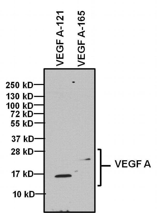
- Submitted by
- Invitrogen Antibodies (provider)
- Main image

- Experimental details
- Western blot analysis of Vascular Endothelial Growth Factor (VEGF) was performed by loading 1 µg of indicated recombinant VEGF isoforms, and 10 µL of PageRuler PlusPrestained Protein Ladder (Product # 26619) per well onto a 4-20% Tris-Glycine polyacrylamide gel. Proteins were transferred to a PVDF membrane (Product # 88518) using the G2 Fast Blotter (Product # 62288) and blocked with 5% Milk/TBST for at least 1 hour at room temperature. VEGF was detected at 14 kD and 19 kD using a VEGF polyclonal antibody (Product # P802) at a dilution of 1:500 in blocking buffer overnight at 4C on a rocking platform, followed by an HRP-conjugated goat anti-rabbit IgG (Fc) secondary antibody (Product # 31463) at a dilution of 1:20,000 for at least 1 hour. Chemiluminescent detection was performed using SuperSignal West Dura (Product # 34076).

Supportive validation
- Submitted by
- Invitrogen Antibodies (provider)
- Main image
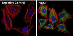
- Experimental details
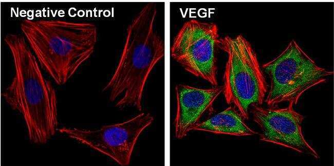
- Immunofluorescent analysis of Vascular Endothelial Growth Factor (VEGF, green) in Hela cells. The cells were fixed with 4% paraformaldehyde, permeabilized with 0.1% Triton X-100 in PBS, and blocked with 3% Blocker BSA (Product # 37525) for 30 minutes at room temperature. Cells were stained with (left panel) or without (right panel) a VEGF polyclonal antibody (Product # P802) at a dilution of 1:40 for at least 1 hour at room temperature, and then incubated with a Dylight 488 goat anti-rabbit IgG secondary antibody at a dilution of 1:1000 for 45 minutes at room temperature. F-actin (both panels, red) was stained by Dylight 554 Phalloidin (Product # 21834) and nuclei (both panels, blue) were stained with DAPI (Product # 46190). Images were taken at 60X magnification.
- Submitted by
- Invitrogen Antibodies (provider)
- Main image

- Experimental details
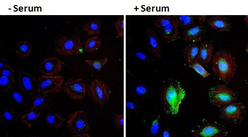
- Immunofluorescent analysis of VEGF (green) in HeLa cells either serum starved (left panel) or treated with serum (right panel) for 30 minutes. Formalin fixed cells were permeabilized with 0.1% Triton X-100 in TBS for 10 minutes at room temperature and blocked with 1% Blocker BSA (Product # 37525) for 15 minutes at room temperature. Cells were probed with a VEGF polyclonal antibody (Product # P802) at a dilution of 1:50 for at least 1 hour at room temperature, washed with PBS, and incubated with DyLight 488 goat anti-rabbit IgG secondary antibody (Product # 35552) at a dilution of 1:400 for 30 minutes at room temperature. F-Actin (red) was stained with DyLight 554 Phalloidin (Product # 21834) and nuclei (blue) were stained with Hoechst 33342 dye (Product # 62249). Images were taken on a Thermo Scientific ArrayScan at 20X magnification.
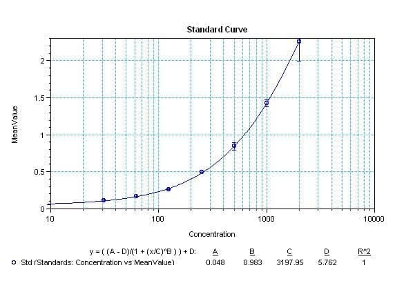
Supportive validation
- Submitted by
- Invitrogen Antibodies (provider)
- Main image

- Experimental details
- Sandwich ELISA analysis of Human VEGF was performed using the Thermo Scientific ELISA Kit (Product # EHVEGF) by coating a blank 96-well microtiter plate with 100 µL per well of Human VEGF moncolonal antibody (Product # M808) in duplicate at 1, 3, 4, 5, 7, and 9 µg/mL in DPBS (Product # 28374) and incubating for 12-18 hours at 4C. The plate was aspirated and blocked with 300 µL per well of 4% BSA and 5% sucrose in DPBS for 1 hour at room temperature. Human VEGF recombinant protein (Product # SVEGF) at 50 µL per well (in addition to 50 µL of sample diluent per well) was added in duplicate at 2000, 1000, 500, 250, 125, 62.5, 31.25, and 0pg/mL for 2 hours at room temperature. The plate was washed with ELISA Wash Buffer (Product # N503) and incubated with 100 µL of Human VEGF biotinylated polyclonal antibody (Product # P802B, biotinylated conjugate of Product # P802) in all applicable wells at 0.5 µg/mL for 1 hour at room temperature. The plate was washed and incubated with 100 µL per well of Streptavidin-HRP (Product # N504) in all test wells at 1:40,000 dilution for 30 minutes at room temperature and then washed and incubated with 100 µL per well of TMB substrate (Product # 34028) for 30 minutes at room temperature in the dark. The plate was stopped with 0.16M sulfuric acid (Product # N600). Absorbances were read on a spectrophotometer at 450-550 nm.