 Explore
 Validate
 Learn
Learn Western blot
 ELISA
ELISAAntibody data
- Antibody Data
- Antigen structure
- References [0]
- Comments [0]
- Validations
- Western blot [2]
- Immunocytochemistry [1]
- Other assay [1]
Submit
Validation data
Reference
Comment
Report error
- Product number
- P802B - Provider product page

- Provider
- Invitrogen Antibodies
- Product name
- VEGF Polyclonal Antibody, Biotin
- Antibody type
- Polyclonal
- Antigen
- Other
- Description
- The VEGF-A monoclonal antibody clone # 16F1 (Product # M808) has successfully been paired as the coating antibody in a sandwich ELISA with detection antibody P802B (biotinylated conjugate of Product # P802). Typical dilutions for sandwich ELISA include 1 µg/mL for coating and 0.125-0.5 µg/mL for detection.
- Reactivity
- Human
- Host
- Rabbit
- Conjugate
- Biotin
- Isotype
- IgG
- Vial size
- 100 µg
- Concentration
- 0.5 mg/mL
- Storage
- -20°C
No comments: Submit comment
Supportive validation
- Submitted by
- Invitrogen Antibodies (provider)
- Main image
- Experimental details
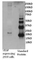
- Western blot analysis of Human VEGF was performed by loading 0. 5 µg of VEGF transfected 293T cell culture supernatant and recombinant human VEGF onto a 4-12% Tris-HCl polyacrylamide gel. Proteins were transferred to a PVDF membrane and blocked with 3% BSA/TBST for at least 1 hour. The membrane was probed with a human VEGF polyclonal antibody (Product # P802) at a dilution of 1 µg/mL overnight at 4°C on a rocking platform, washed in TBS-0.1%Tween 20 and probed with a goat anti- rabbit-HRP secondary antibody (Product # 31460) at a dilution of 1:15,000 for at least one hour. Chemiluminescent detection was performed using SuperSignal West Dura (Product # 34075).
- Conjugate
- Biotin

- Submitted by
- Invitrogen Antibodies (provider)
- Main image

- Experimental details
- Western blot analysis was performed on whole cell extracts (30 µg lysate) of HUVEC (Lane 1), MCF7 (Lane 2), A549 (Lane 3), HAEC (Lane 4) and HeLa (Lane 5). The blot was probed with Rabbit Anti- VEGF Polyclonal Antibody (Product # P802B, 1 µg/mL). A 45 kDa band corresponding to VEGF was observed across the cell lines tested. Known quantity of protein samples were electrophoresed using Novex® NuPAGE® 4-12 % Bis-Tris gel (Product # NP0321BOX), XCell SureLock™ Electrophoresis System (Product # EI0002) and Novex® Sharp Pre-Stained Protein Standard (Product # LC5800). Resolved proteins were then transferred onto a nitrocellulose membrane with iBlot® 2 Dry Blotting System (Product # IB21001). The membrane was probed with the relevant primary antibody following blocking with 5 % skimmed milk. This is followed by incubating the membrane with Poly-HRP Streptavidin (Product # N200, 1:10,000 dilution). Chemiluminescent detection was performed using Pierce™ ECL Western Blotting Substrate (Product # 32106).
- Conjugate
- Biotin

Supportive validation
- Submitted by
- Invitrogen Antibodies (provider)
- Main image

- Experimental details
- Immunofluorescent analysis of VEGF (green) in HeLa cells either serum starved (left panel) or treated with serum (right panel) for 30 minutes. Formalin fixed cells were permeabilized with 0.1% Triton X-100 in TBS for 10 minutes at room temperature and blocked with 1% Blocker BSA (Product # 37525) for 15 minutes at room temperature. Cells were probed with a VEGF polyclonal antibody (Product # P802) at a dilution of 1:50 for at least 1 hour at room temperature, washed with PBS, and incubated with DyLight 488 goat anti-rabbit IgG secondary antibody (Product # 35552) at a dilution of 1:400 for 30 minutes at room temperature. F-Actin (red) was stained with DyLight 554 Phalloidin (Product # 21834) and nuclei (blue) were stained with Hoechst 33342 dye (Product # 62249). Images were taken on a Thermo Scientific ArrayScan at 20X magnification.
- Conjugate
- Biotin

Supportive validation
- Submitted by
- Invitrogen Antibodies (provider)
- Main image

- Experimental details
- Sandwich ELISA analysis of Human VEGF was performed using the Thermo Scientific ELISA Kit (Product # EHVEGF) by coating a blank 96-well microtiter plate with 100 µL per well of Human VEGF moncolonal antibody (Product # M808) in duplicate at 1, 3, 4, 5, 7, and 9 µg/mL in DPBS (Product # 28374) and incubating for 12-18 hours at 4C. The plate was aspirated and blocked with 300 µL per well of 4% BSA and 5% sucrose in DPBS for 1 hour at room temperature. Human VEGF recombinant protein (Product # SVEGF) at 50 µL per well (in addition to 50 µL of sample diluent per well) was added in duplicate at 2000, 1000, 500, 250, 125, 62.5, 31.25, and 0pg/mL for 2 hours at room temperature. The plate was washed with ELISA Wash Buffer (Product # N503) and incubated with 100 µL of Human VEGF biotinylated monoclonal antibody (Product # P802B) in all applicable wells at 0.5 µg/mL for 1 hour at room temperature. The plate was washed and incubated with 100 µL per well of Streptavidin-HRP (Product # N504) in all test wells at 1:40,000 dilution for 30 minutes at room temperature and then washed and incubated with 100 µL per well of TMB substrate (Product # 34028) for 30 minutes at room temperature in the dark. The plate was stopped with 0.16M sulfuric acid (Product # N600). Absorbances were read on a spectrophotometer at 450-550 nm.
- Conjugate
- Biotin
