Explore
 Validate
 Learn
Learn Western blot
 Immunohistochemistry
Antibody data
- Antibody Data
- Antigen structure
- References [66]
- Comments [0]
- Validations
- Immunohistochemistry [1]
Submit
Validation data
Reference
Comment
Report error
- Product number
- PB9071 - Provider product page

- Provider
- Boster Biological Technology
- Product name
- Anti-VEGF/VEGFA Antibody Picoband™
- Antibody type
- Polyclonal
- Description
- Polyclonal antibody for VEGF/VEGFA detection. Host: Rabbit.Size: 100μg/vial. Tested applications: WB, IHC-P, FCM. Reactive species: Human;Mouse;Rat. VEGF/VEGFA information: Molecular Weight: 27042 MW; Subcellular Localization: Secreted . VEGF121 is acidic and freely secreted. VEGF165 is more basic, has heparin-binding properties and, although a signicant proportion remains cell-associated, most is freely secreted. VEGF189 is very basic, it is cell-associated after secretion and is bound avidly by heparin and the extracellular matrix, although it may be released as a soluble form by heparin, heparinase or plasmin; Tissue Specificity: Isoform VEGF189, isoform VEGF165 and isoform VEGF121 are widely expressed. Isoform VEGF206 and isoform VEGF145 are not widely expressed. A higher level expression seen in pituitary tumors as compared to the pituitary gland.
- Reactivity
- Human, Mouse, Rat
- Host
- Rabbit
- Vial size
- 100μg/vial
- Concentration
- Add 0.2ml of distilled water will yield a concentration of 500ug/ml.
- Storage
- At -20°C for one year. After reconstitution, at 4°C for one month. It can also be aliquoted and stored frozen at -20°C for a longer time. Avoid repeated freezing and thawing.
- Handling
- Add 0.2ml of distilled water will yield a concentration of 500ug/ml.
Submitted references Chondroitin sulfate-modified antiangiogenic peptide conjugate induces cell apoptosis via the mitochondria-mediated pathway to perform antitumor activity.
Multifunctional Drug- and AuNRs-Loaded ROS-Responsive Selenium-Containing Polyurethane Nanofibers for Smart Wound Healing.
Low-dose TNF-α promotes angiogenesis of oral squamous cell carcinoma cells via TNFR2/Akt/mTOR axis.
VEGF overexpression in transplanted NSCs promote recovery of neurological function in rats with cerebral ischemia by modulating the Wnt signal transduction pathway.
Chronic intermittent hypobaric hypoxia ameliorates osteoporosis after spinal cord injury through balancing osteoblast and osteoclast activities in rats.
Combinatorial treatment with Silybum marianum essential oil enhances the therapeutic efficacy of a 5-fluorouracil base therapy for hepatocellular carcinoma.
Design, Synthesis and Anti-Melanoma Activity of Novel Annexin V Derivative with β(3)-Integrin Affinity.
Integrin Targeting Enhances the Antimelanoma Effect of Annexin V in Mice.
Inflammation Control and Tumor Growth Inhibition of Ovarian Cancer by Targeting Adhesion Molecules of E-Selectin.
miR-138-5p Inhibits Vascular Mimicry by Targeting the HIF-1α/VEGFA Pathway in Hepatocellular Carcinoma.
Two distinctive types of telocytes in gills of fish: A light, immunohistochemical and ultra-structure study.
A study of diabetes-induced erectile dysfunction treated with human umbilical cord mesenchymal stem cells.
Novel dopamine-modified oxidized sodium alginate hydrogels promote angiogenesis and accelerate healing of chronic diabetic wounds.
Curcumin Derivative Cur20 Attenuated Cerebral Ischemic Injury by Antioxidant Effect and HIF-1α/VEGF/TFEB-Activated Angiogenesis.
An oxidation responsive nano-radiosensitizer increases radiotherapy efficacy by remolding tumor vasculature.
Construction of an AuHQ nano-sensitizer for enhanced radiotherapy efficacy through remolding tumor vasculature.
Hydrogel microfluidic-based liver-on-a-chip: Mimicking the mass transfer and structural features of liver.
Migratory Activities and Stemness Properties of Rodlet Cells.
Synergic effect of PD-1 blockade and endostar on the PI3K/AKT/mTOR-mediated autophagy and angiogenesis in Lewis lung carcinoma mouse model.
Ginsenoside protects against AKI via activation of HIF‑1α and VEGF‑A in the kidney‑brain axis.
MiR-203a-3p inhibits retinal angiogenesis and alleviates proliferative diabetic retinopathy in oxygen-induced retinopathy (OIR) rat model via targeting VEGFA and HIF-1α.
Anti-inflammatory effects of the root, stem and leaf extracts of Chloranthus serratus on adjuvant-induced arthritis in rats.
A Comparison of Ramipril and Bevacizumab to Mitigate Radiation-Induced Brain Necrosis: An Experimental Study.
Recombinant human growth hormone (rhGH) treatment of MKN-45 xenograft mice improves nutrition status and strengthens immune function without promoting tumor growth.
A novel role of HIF-1α/PROX-1/LYVE-1 axis on tissue regeneration after renal ischaemia/reperfusion in mice.
Targeted Delivery of Zoledronate to Tumor-Associated Macrophages for Cancer Immunotherapy.
Effects of CO2 fractional laser on hair growth in C57BL/6 mice and potential underlying mechanisms.
High thoracic sympathetic block improves coronary microcirculation disturbance in rats with chronic heart failure.
Liquiritin from Glycyrrhiza uralensis Attenuating Rheumatoid Arthritis via Reducing Inflammation, Suppressing Angiogenesis, and Inhibiting MAPK Signaling Pathway.
The protective role of intermedin in promoting angiogenesis during renal fibrosis.
MGF E peptide improves anterior cruciate ligament repair by inhibiting hypoxia-induced cell apoptosis and accelerating angiogenesis.
Ginsenoside Rh2 Inhibits Angiogenesis in Prostate Cancer by Targeting CNNM1.
An Experimental Study on Repeated Brief Ischemia in Promoting Sciatic Nerve Repair and Regeneration in Rats.
The earlier, the better: the effects of different administration timepoints of sorafenib in suppressing the carcinogenesis of VEGF in rats.
Vascularization of Lando(®) dermal scaffold in an acute full-thickness skin-defect porcine model.
LncRNA MIAT facilitated BM-MSCs differentiation into endothelial cells and restored erectile dysfunction via targeting miR-200a in a rat model of erectile dysfunction.
Synthetic E-selectin prevents postoperative vascular restenosis by inhibiting nuclear factor κB in rats.
Perineurium-like sheath derived from long-term surviving mesenchymal stem cells confers nerve protection to the injured spinal cord.
The Involvement of β-Catenin/COX-2/VEGF Axis in NMDA-Caused Retinopathy.
Effects of Chronic Exposure to Sodium Arsenite on Expressions of VEGF and VEGFR2 Proteins in the Epididymis of Rats.
Tetrahydrocurcumin induces mesenchymal-epithelial transition and suppresses angiogenesis by targeting HIF-1α and autophagy in human osteosarcoma.
Synergistic effects of G-CSF and bone marrow stromal cells on nerve regeneration with acellular nerve xenografts.
A macrophage-activating, injectable hydrogel to sequester endogenous growth factors for in situ angiogenesis.
Ilexgenin A exerts anti-inflammation and anti-angiogenesis effects through inhibition of STAT3 and PI3K pathways and exhibits synergistic effects with Sorafenib on hepatoma growth.
Effect of tetramethylpyrazine combined with cisplatin on VEGF, KLF4 and ADAMTS1 in Lewis lung cancer mice.
Pioglitazone, a Peroxisome Proliferator-Activated Receptor x03B3; Agonist, Ameliorates Chronic Kidney Disease by Enhancing Antioxidative Capacity and Attenuating Angiogenesis in the Kidney of a 5/6 Nephrectomized Rat Model.
Enhanced antitumor activity and attenuated cardiotoxicity of Epirubicin combined with Paeonol against breast cancer.
A novel multi-target RNAi adenovirus inhibits hepatoma cell proliferation, migration, and induction of angiogenesis.
Buyanghuanwu decoction promotes angiogenesis after cerebral ischemia/reperfusion injury: mechanisms of brain tissue repair.
Lentivirus-mediated PLCγ1 gene short-hairpin RNA suppresses tumor growth and metastasis of human gastric adenocarcinoma.
Overexpression of astrocyte elevated gene-1 (AEG-1) in cervical cancer and its correlation with angiogenesis.
Integration of donor mesenchymal stem cell-derived neuron-like cells into host neural network after rat spinal cord transection.
Study of the mechanism of sonodynamic therapy in a rat glioma model.
Correlations between CT perfusion parameters and vascular endothelial growth factor expression and microvessel density in implanted VX2 lung tumors.
Effects of inhibition of hedgehog signaling on cell growth and migration of uveal melanoma cells.
Preparation, antiangiogenic and antitumoral activities of the chemically sulfated glucan from Phellinus ribis.
Targeted depletion of tumour-associated macrophages by an alendronate-glucomannan conjugate for cancer immunotherapy.
Nuclear factor-κB signaling pathway is involved in phospholipase Cε-regulated proliferation in human renal cell carcinoma cells.
Clinical study of tumor angiogenesis and perfusion imaging using multi-slice spiral computed tomography for breast cancer.
Relationships of uPA and VEGF expression in esophageal cancer and microvascular density with tumorous invasion and metastasis.
Sciatic nerve repair by acellular nerve xenografts implanted with BMSCs in rats xenograft combined with BMSCs.
Effects of Panax notoginoside on the nephropathy in rats with type 1 diabetes mellitus.
Extract of Ginkgo biloba promotes the expression of VEGF following subarachnoid hemorrhage in rats.
Decreased TIP30 expression promotes tumor metastasis in lung cancer.
Induction stage-dependent expression of vascular endothelial growth factor and aquaporin-1 in diethylstilbestrol-treated rat pituitary.
Total saponins of Panax notoginseng protected rabbit iliac artery against balloon endothelial denudation injury.
Li Y, Fu J, Hou H, Tang W, Liu Z, Gao D, Zhao F, Gao X, Sun F, Tan H
International journal of biological macromolecules 2024 Mar;262(Pt 1):129671
International journal of biological macromolecules 2024 Mar;262(Pt 1):129671
Multifunctional Drug- and AuNRs-Loaded ROS-Responsive Selenium-Containing Polyurethane Nanofibers for Smart Wound Healing.
Ahmed W, Li S, Liang M, Kang Y, Liu X, Gao C
ACS biomaterials science & engineering 2024 Jun 10;10(6):3946-3957
ACS biomaterials science & engineering 2024 Jun 10;10(6):3946-3957
Low-dose TNF-α promotes angiogenesis of oral squamous cell carcinoma cells via TNFR2/Akt/mTOR axis.
Li S, Liu W, Liu J, Yang Z, Zhang L, Nie F, Yang P, Guo H, Yang C
Oral diseases 2024 Jul;30(5):3004-3017
Oral diseases 2024 Jul;30(5):3004-3017
VEGF overexpression in transplanted NSCs promote recovery of neurological function in rats with cerebral ischemia by modulating the Wnt signal transduction pathway.
Zhu Y, Liu R, Zhao X, Kang C, Yang D, Ge G
Neuroscience letters 2024 Feb 28;824:137668
Neuroscience letters 2024 Feb 28;824:137668
Chronic intermittent hypobaric hypoxia ameliorates osteoporosis after spinal cord injury through balancing osteoblast and osteoclast activities in rats.
Zhang L, Yin Y, Guo J, Jin L, Hou Z
Frontiers in endocrinology 2023;14:1035186
Frontiers in endocrinology 2023;14:1035186
Combinatorial treatment with Silybum marianum essential oil enhances the therapeutic efficacy of a 5-fluorouracil base therapy for hepatocellular carcinoma.
MasodKhooy MJ, Farasat M, Salehi Salmi M, Mirzaei H
Phytotherapy research : PTR 2023 May;37(5):1968-1985
Phytotherapy research : PTR 2023 May;37(5):1968-1985
Design, Synthesis and Anti-Melanoma Activity of Novel Annexin V Derivative with β(3)-Integrin Affinity.
Zhu J, Li W, Jing J
International journal of molecular sciences 2023 Jul 5;24(13)
International journal of molecular sciences 2023 Jul 5;24(13)
Integrin Targeting Enhances the Antimelanoma Effect of Annexin V in Mice.
Zhu J, Li X, Gao W, Jing J
International journal of molecular sciences 2023 Feb 15;24(4)
International journal of molecular sciences 2023 Feb 15;24(4)
Inflammation Control and Tumor Growth Inhibition of Ovarian Cancer by Targeting Adhesion Molecules of E-Selectin.
Yang B, Yin S, Zhou Z, Huang L, Xi M
Cancers 2023 Apr 4;15(7)
Cancers 2023 Apr 4;15(7)
miR-138-5p Inhibits Vascular Mimicry by Targeting the HIF-1α/VEGFA Pathway in Hepatocellular Carcinoma.
Liu H, Tang T, Hu X, Tan W, Zhou P, Zhang H, Liu Y, Chen C, Yang M, Zhou M, Xuan S, Cheng B, Yin W, Lin J
Journal of immunology research 2022;2022:7318950
Journal of immunology research 2022;2022:7318950
Two distinctive types of telocytes in gills of fish: A light, immunohistochemical and ultra-structure study.
Soliman SA, Sobh A, Ali LA, Abd-Elhafeez HH
Microscopy research and technique 2022 Nov;85(11):3653-3663
Microscopy research and technique 2022 Nov;85(11):3653-3663
A study of diabetes-induced erectile dysfunction treated with human umbilical cord mesenchymal stem cells.
Wang S, Zhang A, Liu K, Pan Y, Kang J, Niu S, Song Y, Zhang Z, Li Y, Liu L, Liu X
Andrologia 2022 Aug;54(7):e14440
Andrologia 2022 Aug;54(7):e14440
Novel dopamine-modified oxidized sodium alginate hydrogels promote angiogenesis and accelerate healing of chronic diabetic wounds.
Chi J, Li A, Zou M, Wang S, Liu C, Hu R, Jiang Z, Liu W, Sun R, Han B
International journal of biological macromolecules 2022 Apr 1;203:492-504
International journal of biological macromolecules 2022 Apr 1;203:492-504
Curcumin Derivative Cur20 Attenuated Cerebral Ischemic Injury by Antioxidant Effect and HIF-1α/VEGF/TFEB-Activated Angiogenesis.
Zhang R, Zhao T, Zheng B, Zhang Y, Li X, Zhang F, Cen J, Duan S
Frontiers in pharmacology 2021;12:648107
Frontiers in pharmacology 2021;12:648107
An oxidation responsive nano-radiosensitizer increases radiotherapy efficacy by remolding tumor vasculature.
Wang X, Niu X, Sha W, Feng X, Yu L, Zhang Z, Wang W, Yuan Z
Biomaterials science 2021 Sep 14;9(18):6308-6324
Biomaterials science 2021 Sep 14;9(18):6308-6324
Construction of an AuHQ nano-sensitizer for enhanced radiotherapy efficacy through remolding tumor vasculature.
Wang X, Niu X, Zhang X, Zhang Z, Gao X, Wang W, Yuan Z
Journal of materials chemistry. B 2021 Jun 3;9(21):4365-4379
Journal of materials chemistry. B 2021 Jun 3;9(21):4365-4379
Hydrogel microfluidic-based liver-on-a-chip: Mimicking the mass transfer and structural features of liver.
Meng Q, Wang Y, Li Y, Shen C
Biotechnology and bioengineering 2021 Feb;118(2):612-621
Biotechnology and bioengineering 2021 Feb;118(2):612-621
Migratory Activities and Stemness Properties of Rodlet Cells.
Abd-Elhafeez HH, Abou-Elhamd AS, Abdo W, Soliman SA
Microscopy and microanalysis : the official journal of Microscopy Society of America, Microbeam Analysis Society, Microscopical Society of Canada 2020 Oct;26(5):1035-1052
Microscopy and microanalysis : the official journal of Microscopy Society of America, Microbeam Analysis Society, Microscopical Society of Canada 2020 Oct;26(5):1035-1052
Synergic effect of PD-1 blockade and endostar on the PI3K/AKT/mTOR-mediated autophagy and angiogenesis in Lewis lung carcinoma mouse model.
Wu J, Zhao X, Sun Q, Jiang Y, Zhang W, Luo J, Li Y
Biomedicine & pharmacotherapy = Biomedecine & pharmacotherapie 2020 May;125:109746
Biomedicine & pharmacotherapy = Biomedecine & pharmacotherapie 2020 May;125:109746
Ginsenoside protects against AKI via activation of HIF‑1α and VEGF‑A in the kidney‑brain axis.
Mao H, Jiang C, Xu L, Chen D, Liu H, Xu Y, Ma K, Wang M
International journal of molecular medicine 2020 Mar;45(3):939-946
International journal of molecular medicine 2020 Mar;45(3):939-946
MiR-203a-3p inhibits retinal angiogenesis and alleviates proliferative diabetic retinopathy in oxygen-induced retinopathy (OIR) rat model via targeting VEGFA and HIF-1α.
Han N, Xu H, Yu N, Wu Y, Yu L
Clinical and experimental pharmacology & physiology 2020 Jan;47(1):85-94
Clinical and experimental pharmacology & physiology 2020 Jan;47(1):85-94
Anti-inflammatory effects of the root, stem and leaf extracts of Chloranthus serratus on adjuvant-induced arthritis in rats.
Sun S, Li S, Du Y, Wu C, Zhang M, Li J, Zhang X
Pharmaceutical biology 2020 Dec;58(1):528-537
Pharmaceutical biology 2020 Dec;58(1):528-537
A Comparison of Ramipril and Bevacizumab to Mitigate Radiation-Induced Brain Necrosis: An Experimental Study.
Erpolat OP, Demircan NV, Sarıbas GS, Kuzucu P, Senturk E, Elmas C, Borcek A, Kurt G
World neurosurgery 2020 Dec;144:e210-e220
World neurosurgery 2020 Dec;144:e210-e220
Recombinant human growth hormone (rhGH) treatment of MKN-45 xenograft mice improves nutrition status and strengthens immune function without promoting tumor growth.
Wei L, Chang J, Han Z, Wang R, Song L
PloS one 2019;14(1):e0210613
PloS one 2019;14(1):e0210613
A novel role of HIF-1α/PROX-1/LYVE-1 axis on tissue regeneration after renal ischaemia/reperfusion in mice.
Meng F
Archives of physiology and biochemistry 2019 Oct;125(4):321-331
Archives of physiology and biochemistry 2019 Oct;125(4):321-331
Targeted Delivery of Zoledronate to Tumor-Associated Macrophages for Cancer Immunotherapy.
Zang X, Zhang X, Hu H, Qiao M, Zhao X, Deng Y, Chen D
Molecular pharmaceutics 2019 May 6;16(5):2249-2258
Molecular pharmaceutics 2019 May 6;16(5):2249-2258
Effects of CO2 fractional laser on hair growth in C57BL/6 mice and potential underlying mechanisms.
Zhuo FL, Li LF, Cai LQ, Huang Y
Chinese medical journal 2019 May 20;132(10):1257-1260
Chinese medical journal 2019 May 20;132(10):1257-1260
High thoracic sympathetic block improves coronary microcirculation disturbance in rats with chronic heart failure.
Sun G, Liu F, Xiu C
Microvascular research 2019 Mar;122:94-100
Microvascular research 2019 Mar;122:94-100
Liquiritin from Glycyrrhiza uralensis Attenuating Rheumatoid Arthritis via Reducing Inflammation, Suppressing Angiogenesis, and Inhibiting MAPK Signaling Pathway.
Zhai KF, Duan H, Cui CY, Cao YY, Si JL, Yang HJ, Wang YC, Cao WG, Gao GZ, Wei ZJ
Journal of agricultural and food chemistry 2019 Mar 13;67(10):2856-2864
Journal of agricultural and food chemistry 2019 Mar 13;67(10):2856-2864
The protective role of intermedin in promoting angiogenesis during renal fibrosis.
Dong H, Zhou Y, Wang Y, Zhou Q, Zhang Y, Gan X, Luo Y, Li R
Gene 2019 Mar 10;688:34-43
Gene 2019 Mar 10;688:34-43
MGF E peptide improves anterior cruciate ligament repair by inhibiting hypoxia-induced cell apoptosis and accelerating angiogenesis.
Sha Y, Yang L, Lv Y
Journal of cellular physiology 2019 Jun;234(6):8846-8861
Journal of cellular physiology 2019 Jun;234(6):8846-8861
Ginsenoside Rh2 Inhibits Angiogenesis in Prostate Cancer by Targeting CNNM1.
Huang Y, Huang H, Han Z, Li W, Mai Z, Yuan R
Journal of nanoscience and nanotechnology 2019 Apr 1;19(4):1942-1950
Journal of nanoscience and nanotechnology 2019 Apr 1;19(4):1942-1950
An Experimental Study on Repeated Brief Ischemia in Promoting Sciatic Nerve Repair and Regeneration in Rats.
Zhou XB, Zou DX, Gu W, Wang D, Feng JS, Wang JY, Zhou JL
World neurosurgery 2018 Jun;114:e11-e21
World neurosurgery 2018 Jun;114:e11-e21
The earlier, the better: the effects of different administration timepoints of sorafenib in suppressing the carcinogenesis of VEGF in rats.
Li N, Chen B, Lin R, Liu N, Dai HT, Tang KY, Yang JY, Huang YH
Cancer chemotherapy and pharmacology 2018 Jan;81(1):207-216
Cancer chemotherapy and pharmacology 2018 Jan;81(1):207-216
Vascularization of Lando(®) dermal scaffold in an acute full-thickness skin-defect porcine model.
Qiu X, Wang J, Wang G, Wen H
Journal of plastic surgery and hand surgery 2018 Aug;52(4):204-209
Journal of plastic surgery and hand surgery 2018 Aug;52(4):204-209
LncRNA MIAT facilitated BM-MSCs differentiation into endothelial cells and restored erectile dysfunction via targeting miR-200a in a rat model of erectile dysfunction.
Wang H, Ding XG, Yang JJ, Li SW, Zheng H, Gu CH, Jia ZK, Li L
European journal of cell biology 2018 Apr;97(3):180-189
European journal of cell biology 2018 Apr;97(3):180-189
Synthetic E-selectin prevents postoperative vascular restenosis by inhibiting nuclear factor κB in rats.
Liu J, Liu Z, Hu X, Zhang Y, Zhang S
Molecular medicine reports 2018 Apr;17(4):5065-5073
Molecular medicine reports 2018 Apr;17(4):5065-5073
Perineurium-like sheath derived from long-term surviving mesenchymal stem cells confers nerve protection to the injured spinal cord.
Ma YH, Zeng X, Qiu XC, Wei QS, Che MT, Ding Y, Liu Z, Wu GH, Sun JH, Pang M, Rong LM, Liu B, Aljuboori Z, Han I, Ling EA, Zeng YS
Biomaterials 2018 Apr;160:37-55
Biomaterials 2018 Apr;160:37-55
The Involvement of β-Catenin/COX-2/VEGF Axis in NMDA-Caused Retinopathy.
Ning D, Zhang WK, Tian H, Li XJ, Liu M, Li YS, Tang HB
Journal of ophthalmology 2017;2017:9760501
Journal of ophthalmology 2017;2017:9760501
Effects of Chronic Exposure to Sodium Arsenite on Expressions of VEGF and VEGFR2 Proteins in the Epididymis of Rats.
Yan-Ping D, Xiao-Qin G, Xiao Ping M, Ying Quan Y
BioMed research international 2017;2017:2597256
BioMed research international 2017;2017:2597256
Tetrahydrocurcumin induces mesenchymal-epithelial transition and suppresses angiogenesis by targeting HIF-1α and autophagy in human osteosarcoma.
Zhang Y, Liu Y, Zou J, Yan L, Du W, Zhang Y, Sun H, Lu P, Geng S, Gu R, Zhang H, Bi Z
Oncotarget 2017 Oct 31;8(53):91134-91149
Oncotarget 2017 Oct 31;8(53):91134-91149
Synergistic effects of G-CSF and bone marrow stromal cells on nerve regeneration with acellular nerve xenografts.
Jia H, Wang Y, Wang T, Dong Y, Li WL, Li JP, Ma WZ, Tong XJ, He ZY
Synapse (New York, N.Y.) 2017 Jul;71(7)
Synapse (New York, N.Y.) 2017 Jul;71(7)
A macrophage-activating, injectable hydrogel to sequester endogenous growth factors for in situ angiogenesis.
Feng Y, Li Q, Wu D, Niu Y, Yang C, Dong L, Wang C
Biomaterials 2017 Jul;134:128-142
Biomaterials 2017 Jul;134:128-142
Ilexgenin A exerts anti-inflammation and anti-angiogenesis effects through inhibition of STAT3 and PI3K pathways and exhibits synergistic effects with Sorafenib on hepatoma growth.
Yang H, Wang J, Fan JH, Zhang YQ, Zhao JX, Dai XJ, Liu Q, Shen YJ, Liu C, Sun WD, Sun Y
Toxicology and applied pharmacology 2017 Jan 15;315:90-101
Toxicology and applied pharmacology 2017 Jan 15;315:90-101
Effect of tetramethylpyrazine combined with cisplatin on VEGF, KLF4 and ADAMTS1 in Lewis lung cancer mice.
Tang JH, Zhang HM, Zhang ZH, Zhang XL
Asian Pacific journal of tropical medicine 2017 Aug;10(8):813-818
Asian Pacific journal of tropical medicine 2017 Aug;10(8):813-818
Pioglitazone, a Peroxisome Proliferator-Activated Receptor x03B3; Agonist, Ameliorates Chronic Kidney Disease by Enhancing Antioxidative Capacity and Attenuating Angiogenesis in the Kidney of a 5/6 Nephrectomized Rat Model.
Sun L, Yuan Q, Xu T, Yao L, Feng J, Ma J, Wang L, Lu C, Wang D
Cellular physiology and biochemistry : international journal of experimental cellular physiology, biochemistry, and pharmacology 2016;38(5):1831-40
Cellular physiology and biochemistry : international journal of experimental cellular physiology, biochemistry, and pharmacology 2016;38(5):1831-40
Enhanced antitumor activity and attenuated cardiotoxicity of Epirubicin combined with Paeonol against breast cancer.
Wu J, Xue X, Zhang B, Cao H, Kong F, Jiang W, Li J, Sun D, Guo R
Tumour biology : the journal of the International Society for Oncodevelopmental Biology and Medicine 2016 Sep;37(9):12301-12313
Tumour biology : the journal of the International Society for Oncodevelopmental Biology and Medicine 2016 Sep;37(9):12301-12313
A novel multi-target RNAi adenovirus inhibits hepatoma cell proliferation, migration, and induction of angiogenesis.
Huang M, Li G, Pan T, Cheng Y, Ren W, Jia W, Ma J, Xu G
Oncotarget 2016 Sep 6;7(36):57705-57713
Oncotarget 2016 Sep 6;7(36):57705-57713
Buyanghuanwu decoction promotes angiogenesis after cerebral ischemia/reperfusion injury: mechanisms of brain tissue repair.
Zhang ZQ, Song JY, Jia YQ, Zhang YK
Neural regeneration research 2016 Mar;11(3):435-40
Neural regeneration research 2016 Mar;11(3):435-40
Lentivirus-mediated PLCγ1 gene short-hairpin RNA suppresses tumor growth and metastasis of human gastric adenocarcinoma.
Zhang B, Wang F, Dai L, Cai H, Zhan Y, Gang S, Hu T, Xia C, Zhang B
Oncotarget 2016 Feb 16;7(7):8043-54
Oncotarget 2016 Feb 16;7(7):8043-54
Overexpression of astrocyte elevated gene-1 (AEG-1) in cervical cancer and its correlation with angiogenesis.
Yu JQ, Zhou Q, Zhu H, Zheng FY, Chen ZW
Asian Pacific journal of cancer prevention : APJCP 2015;16(6):2277-81
Asian Pacific journal of cancer prevention : APJCP 2015;16(6):2277-81
Integration of donor mesenchymal stem cell-derived neuron-like cells into host neural network after rat spinal cord transection.
Zeng X, Qiu XC, Ma YH, Duan JJ, Chen YF, Gu HY, Wang JM, Ling EA, Wu JL, Wu W, Zeng YS
Biomaterials 2015 Jun;53:184-201
Biomaterials 2015 Jun;53:184-201
Study of the mechanism of sonodynamic therapy in a rat glioma model.
Song D, Yue W, Li Z, Li J, Zhao J, Zhang N
OncoTargets and therapy 2014;7:1801-10
OncoTargets and therapy 2014;7:1801-10
Correlations between CT perfusion parameters and vascular endothelial growth factor expression and microvessel density in implanted VX2 lung tumors.
Ling S, Deng D, Mo Y, Zhang X, Guan X, Wei Q
Cell biochemistry and biophysics 2014 Sep;70(1):629-33
Cell biochemistry and biophysics 2014 Sep;70(1):629-33
Effects of inhibition of hedgehog signaling on cell growth and migration of uveal melanoma cells.
Duan F, Lin M, Li C, Ding X, Qian G, Zhang H, Ge S, Fan X, Li J
Cancer biology & therapy 2014 May;15(5):544-59
Cancer biology & therapy 2014 May;15(5):544-59
Preparation, antiangiogenic and antitumoral activities of the chemically sulfated glucan from Phellinus ribis.
Liu Y, Liu Y, Jiang H, Xu L, Cheng Y, Wang PG, Wang F
Carbohydrate polymers 2014 Jun 15;106:42-8
Carbohydrate polymers 2014 Jun 15;106:42-8
Targeted depletion of tumour-associated macrophages by an alendronate-glucomannan conjugate for cancer immunotherapy.
Zhan X, Jia L, Niu Y, Qi H, Chen X, Zhang Q, Zhang J, Wang Y, Dong L, Wang C
Biomaterials 2014 Dec;35(38):10046-57
Biomaterials 2014 Dec;35(38):10046-57
Nuclear factor-κB signaling pathway is involved in phospholipase Cε-regulated proliferation in human renal cell carcinoma cells.
Du HF, Ou LP, Song XD, Fan YR, Yang X, Tan B, Quan Z, Luo CL, Wu XH
Molecular and cellular biochemistry 2014 Apr;389(1-2):265-75
Molecular and cellular biochemistry 2014 Apr;389(1-2):265-75
Clinical study of tumor angiogenesis and perfusion imaging using multi-slice spiral computed tomography for breast cancer.
Xu N, Lei Z, Li XL, Zhang J, Li C, Feng GQ, Li DN, Liu JY, Wei Q, Bian TT, Zou TY
Asian Pacific journal of cancer prevention : APJCP 2013;14(1):429-33
Asian Pacific journal of cancer prevention : APJCP 2013;14(1):429-33
Relationships of uPA and VEGF expression in esophageal cancer and microvascular density with tumorous invasion and metastasis.
Jiang JT, Zhang LF, Zhou B, Zhang SQ, Li SM, Zhang W, Zhang J, Qiao Z, Kong RR, Ma YF, Chen S
Asian Pacific journal of cancer prevention : APJCP 2012;13(7):3379-83
Asian Pacific journal of cancer prevention : APJCP 2012;13(7):3379-83
Sciatic nerve repair by acellular nerve xenografts implanted with BMSCs in rats xenograft combined with BMSCs.
Jia H, Wang Y, Tong XJ, Liu GB, Li Q, Zhang LX, Sun XH
Synapse (New York, N.Y.) 2012 Mar;66(3):256-69
Synapse (New York, N.Y.) 2012 Mar;66(3):256-69
Effects of Panax notoginoside on the nephropathy in rats with type 1 diabetes mellitus.
Tu QN, Dong H, Lu FE
Chinese journal of integrative medicine 2011 Aug;17(8):612-5
Chinese journal of integrative medicine 2011 Aug;17(8):612-5
Extract of Ginkgo biloba promotes the expression of VEGF following subarachnoid hemorrhage in rats.
Sun BL, Hu DM, Yuan H, Ye WJ, Wang XC, Xia ZL, Zhang SM, Wang LX
The International journal of neuroscience 2009;119(7):995-1005
The International journal of neuroscience 2009;119(7):995-1005
Decreased TIP30 expression promotes tumor metastasis in lung cancer.
Tong X, Li K, Luo Z, Lu B, Liu X, Wang T, Pang M, Liang B, Tan M, Wu M, Zhao J, Guo Y
The American journal of pathology 2009 May;174(5):1931-9
The American journal of pathology 2009 May;174(5):1931-9
Induction stage-dependent expression of vascular endothelial growth factor and aquaporin-1 in diethylstilbestrol-treated rat pituitary.
Zhao W, Shen H, Yuan F, Li G, Sun Y, Shi Z, Zhang Y, Wang Z
European journal of histochemistry : EJH 2009 Mar 31;53(1):e7
European journal of histochemistry : EJH 2009 Mar 31;53(1):e7
Total saponins of Panax notoginseng protected rabbit iliac artery against balloon endothelial denudation injury.
Chen SW, Li XH, Ye KH, Jiang ZF, Ren XD
Acta pharmacologica Sinica 2004 Sep;25(9):1151-6
Acta pharmacologica Sinica 2004 Sep;25(9):1151-6
No comments: Submit comment
Supportive validation
- Submitted by
- Boster Biological Technology (provider)
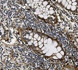
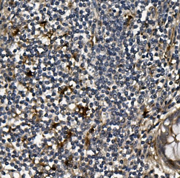
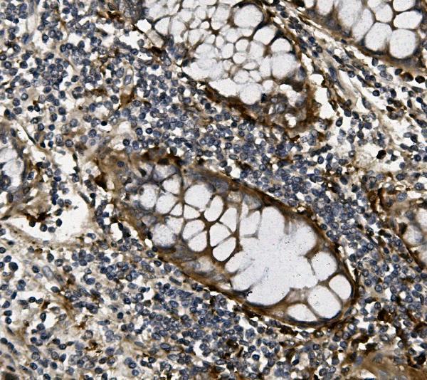
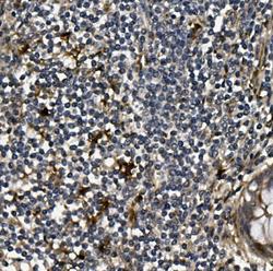
- Main image
- Experimental details
- IHC analysis of VEGF using anti-VEGF antibody (PB9071). VEGF was detected in paraffin-embedded section of human rectal cancer tissue. Heat mediated antigen retrieval was performed in EDTA buffer (pH8.0, epitope retrieval solution). The tissue section was blocked with 10% goat serum. The tissue section was then incubated with 1μg/ml rabbit anti-VEGF Antibody (PB9071) overnight at 4°C. Biotinylated goat anti-rabbit IgG was used as secondary antibody and incubated for 30 minutes at 37°C. The tissue section was developed using Strepavidin-Biotin-Complex (SABC)(Catalog # SA1022) with DAB as the chromogen.
- Additional image