 Explore
 Validate
 Learn
Learn Western blot
Antibody data
- Antibody Data
- Antigen structure
- References [6]
- Comments [0]
- Validations
- Western blot [2]
- Immunocytochemistry [1]
Submit
Validation data
Reference
Comment
Report error
- Product number
- MA5-12949 - Provider product page

- Provider
- Invitrogen Antibodies
- Product name
- Anti-VEGF Monoclonal Antibody (5C3.F8)
- Antibody type
- Monoclonal
- Antigen
- Other
- Description
- MA5-12949 targets Vascular Endothelial Growth Factor in WB and ICC/IF applications and shows reactivity with Human samples.
- Reactivity
- Human
- Host
- Mouse
- Isotype
- IgG
- Antibody clone number
- 5C3.F8
- Vial size
- 500 µL
- Concentration
- 0.2 mg/mL
- Storage
- 4° C
Submitted references The role of stem cell factor and c-KIT in keloid pathogenesis: do tyrosine kinase inhibitors have a potential therapeutic role?
Synergistic effect of celecoxib on 5-fluorouracil-induced apoptosis in hepatocellular carcinoma patients.
Anti-human vascular endothelial growth factor (VEGF) antibody selection for immunohistochemical staining of proliferating blood vessels.
In vitro angiogenesis by human umbilical vein endothelial cells (HUVEC) induced by three-dimensional co-culture with glioblastoma cells.
Brain-specific angiogenesis inhibitor 2 regulates VEGF through GABP that acts as a transcriptional repressor.
The molecular mechanism underlying angiogenesis in hepatocellular carcinoma: the imbalance activation of signaling pathways.
Mukhopadhyay A, Do DV, Ong CT, Khoo YT, Masilamani J, Chan SY, Vincent AS, Wong PK, Lim CP, Cao X, Lim IJ, Phan TT
The British journal of dermatology 2011 Feb;164(2):372-86
The British journal of dermatology 2011 Feb;164(2):372-86
Synergistic effect of celecoxib on 5-fluorouracil-induced apoptosis in hepatocellular carcinoma patients.
Bassiouny AR, Zaky A, Neenaa HM
Annals of hepatology 2010 Oct-Dec;9(4):410-8
Annals of hepatology 2010 Oct-Dec;9(4):410-8
Anti-human vascular endothelial growth factor (VEGF) antibody selection for immunohistochemical staining of proliferating blood vessels.
van der Loos CM, Meijer-Jorna LB, Broekmans ME, Ploegmakers HP, Teeling P, de Boer OJ, van der Wal AC
The journal of histochemistry and cytochemistry : official journal of the Histochemistry Society 2010 Feb;58(2):109-18
The journal of histochemistry and cytochemistry : official journal of the Histochemistry Society 2010 Feb;58(2):109-18
In vitro angiogenesis by human umbilical vein endothelial cells (HUVEC) induced by three-dimensional co-culture with glioblastoma cells.
Chen Z, Htay A, Dos Santos W, Gillies GT, Fillmore HL, Sholley MM, Broaddus WC
Journal of neuro-oncology 2009 Apr;92(2):121-8
Journal of neuro-oncology 2009 Apr;92(2):121-8
Brain-specific angiogenesis inhibitor 2 regulates VEGF through GABP that acts as a transcriptional repressor.
Jeong BC, Kim MY, Lee JH, Kee HJ, Kho DH, Han KE, Qian YR, Kim JK, Kim KK
FEBS letters 2006 Jan 23;580(2):669-76
FEBS letters 2006 Jan 23;580(2):669-76
The molecular mechanism underlying angiogenesis in hepatocellular carcinoma: the imbalance activation of signaling pathways.
Zhao ZC, Zheng SS, Wan YL, Jia CK, Xie HY
Hepatobiliary & pancreatic diseases international : HBPD INT 2003 Nov;2(4):529-36
Hepatobiliary & pancreatic diseases international : HBPD INT 2003 Nov;2(4):529-36
No comments: Submit comment
Supportive validation
- Submitted by
- Invitrogen Antibodies (provider)
- Main image

- Experimental details
- Western blot of Vascular Endothelial Growth Factor using Vascular Endothelial Growth Factor Monoclonal Antibody (Product # MA5-12949) on VEGF Cells.

- Submitted by
- Invitrogen Antibodies (provider)
- Main image
- Experimental details
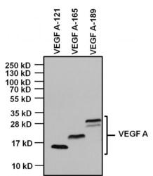
- Western blot analysis of Vascular Endothelial Growth Factor (VEGF) was performed by loading 1ug of indicated recombinant VEGF isoforms, and 10ul of PageRuler PlusPrestained Protein Ladder (Product # 26619) per well onto a 4-20% Tris-Glycine polyacrylamide gel. Proteins were transferred to a PVDF membrane (Product # 88518) using the G2 Fast Blotter (Product # 62288) and blocked with 5% Milk/TBST for at least 1 hour at room temperature. VEGF was detected at 14kD, 19kD and 22-30 kD using a VEGF monoclonal antibody (Product # MA5-12949) at a concentration of 1 µg/mL in blocking buffer overnight at 4C on a rocking platform, followed by an HRP-conjugated goat anti-mouse IgG (Fc) secondary antibody (Product # 31437) at a dilution of 1:20,000 for at least 1 hour. Chemiluminescent detection was performed using SuperSignal West Dura (Product # 34076).

Supportive validation
- Submitted by
- Invitrogen Antibodies (provider)
- Main image

- Experimental details
- Immunofluorescent analysis of Vascular Endothelial Growth Factor (VEGF, green) in Hela cells. The cells were fixed with 4% paraformaldehyde, permeabilized with 0.1% Triton X-100 in PBS, and blocked with 3% Blocker BSA (Product # 37525) for 30 minutes at room temperature. Cells were stained with (left panel) or without (right panel) a VEGF polyclonal antibody (Product # MA5-13182) at a dilution of 1:20 for at least 1 hour at room temperature, and then incubated with a Dylight 488 goat anti-mouse IgG secondary antibody at a dilution of 1:1000 for 45 minutes at room temperature. F-actin (both panels, red) was stained by Dylight 554 Phalloidin (Product # 21834) and nuclei (both panels, blue) were stained with DAPI (Product # 46190). Images were taken at 60X magnification.
