Explore
 Validate
 Learn
Learn Western blot
 Immunohistochemistry
Antibody data
- Antibody Data
- Antigen structure
- References [2]
- Comments [0]
- Validations
- Western blot [2]
- Immunocytochemistry [1]
Submit
Validation data
Reference
Comment
Report error
- Product number
- NB600-548 - Provider product page

- Provider
- Novus Biologicals
- Proper citation
- Novus Cat#NB600-548, RRID:AB_10002448
- Product name
- Mouse Monoclonal VEGF Antibody
- Antibody type
- Monoclonal
- Description
- Protein G purified. VEGF (5C3
- Reactivity
- Human, Mouse
- Host
- Mouse
- Isotype
- IgG
- Vial size
- 500 uL
- Concentration
- 0.2 mg/ml
- Storage
- Store at 4C. Do not freeze.
Submitted references In vitro and in vivo evaluation of N,N,N-trimethylphytosphingosine-iodide (TMP) in liposomes for the treatment of angiogenesis and metastasis.
Analysis of mTOR inhibition-involved pathway in ovarian clear cell adenocarcinoma.
Song CK, Lee JH, Jahn A, Choi MJ, Namgoong SK, Hong SS, Chong S, Shim CK, Chung SJ, Kim DD
International journal of pharmaceutics 2012 Sep 15;434(1-2):191-8
International journal of pharmaceutics 2012 Sep 15;434(1-2):191-8
Analysis of mTOR inhibition-involved pathway in ovarian clear cell adenocarcinoma.
Harasawa M, Yasuda M, Hirasawa T, Miyazawa M, Shida M, Muramatsu T, Douguchi K, Matsui N, Takekoshi S, Kajiwara H, Yoshiyuki Osamura R, Mikami M
Acta histochemica et cytochemica 2011 Apr 28;44(2):113-8
Acta histochemica et cytochemica 2011 Apr 28;44(2):113-8
No comments: Submit comment
Supportive validation
- Submitted by
- Novus Biologicals (provider)
- Main image

- Experimental details
- Western Blot: VEGF Antibody (5C3.F8) [NB600-548] - Analysis of VEGF from Human Cells in a reducing gel.

- Submitted by
- Novus Biologicals (provider)
- Main image

- Experimental details
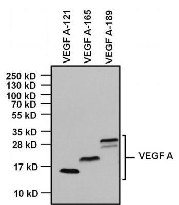
- Western Blot: VEGF Antibody (5C3.F8) [NB600-548] - Analysis of 1ug of indicated recombinant VEGF isoforms, and 10ul of PageRuler PlusPrestained Protein Ladder per well onto a 4-20% Tris-Glycine polyacrylamide gel.
Supportive validation
- Submitted by
- Novus Biologicals (provider)
- Main image

- Experimental details
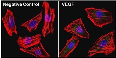
- Immunocytochemistry/Immunofluorescence: VEGF Antibody (5C3.F8) [NB600-548] - Analysis of Vascular Endothelial Growth Factor (VEGF, green) in Hela cells. The cells were fixed with 4% paraformaldehyde, permeabilized with 0.1% Triton X-100 in PBS, and blocked with 3% Blocker BSA for 30 minutes at room temperature. Cells were stained with (left panel) or without (right panel) a VEGF polyclonal antibody at a dilution of 1:20 for at least 1 hour at room temperature, and then incubated with a Dylight 488 goat anti-mouse IgG secondary antibody at a dilution of 1:1000 for 45 minutes at room temperature. F-actin (both panels, red) was stained by Dylight 554 Phalloidin and nuclei (both panels, blue) were stained with DAPI.