Explore
 Validate
 Learn
Learn Western blot
Antibody data
- Antibody Data
- Antigen structure
- References [0]
- Comments [0]
- Validations
- Western blot [1]
- Immunohistochemistry [3]
- Flow cytometry [1]
Submit
Validation data
Reference
Comment
Report error
- Product number
- 10-7007 - Provider product page

- Provider
- ABEOMICS Inc.
- Product name
- Anti-MBD1 Antibody
- Antibody type
- Monoclonal
- Description
- MBD1 (Methyl-CpG-Binding Domain Protein 1) protein is a primary candidate for the readout of DNA methylation as it recruits chromatin remodelers, histone deacetylases and methylases to methylated DNA associated with gene repression. This protein, a member of a transcriptional repressor family MBD, is predominantly expressed in neurons. MBD protein binding requires both functional MBD domains and methyl-CpGs; however, some MBD proteins also bind unmethylated DNA and active regulatory regions via alternative regulatory domains or interaction with the NuRD/Mi-2 (Nucleosome Remodeling Deacetylase) complex members. The CXXC3 domain of MBD1 makes it a unique member of the MBD family due to its affinity to unmethylated DNA. MBD1 acts as an epigenetic regulator via different mechanisms, such as the formation of the MCAF1/MBD1/SETDB1 complex or the MBD1-HDAC3 complex. It also plays an important role in disease progression, contributes to the drug resistance of PC cells; however, the mechanism underlying the drug resistance endowed by MBD1 remains unknown.
- Reactivity
- Human
- Host
- Mouse
- Conjugate
- Unconjugated
- Antigen sequence
A partial length recombinant MBD1 p
rotein (amino acids 291-586) was us
ed as the immunogen for this antibo
dy.- Isotype
- IgG
- Antibody clone number
- ABM15H2
- Vial size
- 100 µg
- Concentration
- 0.5 mg/ml
- Storage
- Store the antibody at 4°C, stable for 6 months. For long-term storage, store at -20°C. Avoid repeat freez thawing
No comments: Submit comment
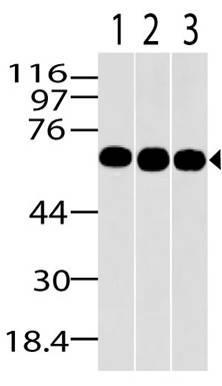
Supportive validation
- Submitted by
- ABEOMICS Inc. (provider)
- Main image

- Experimental details
- Western blot analysis of MBD1. Anti-MBD1 antibody (Clone: ABM15H2) was used at 2 µg/ml on HepG2, MCF7 and A431 lysates.
- Protocol
- Protocol
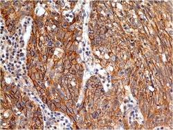
Supportive validation
- Submitted by
- ABEOMICS Inc. (provider)
- Main image
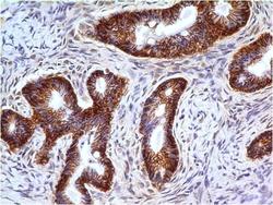
- Experimental details
- Immunohistochemical analysis of MBD1 in Cystadenocarcinoma of ovary using MBD1 antibody (Clone: ABM15H2) at 5 µg/ml.
- Protocol
- Protocol

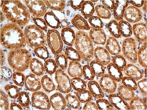
- Submitted by
- ABEOMICS Inc. (provider)
- Main image
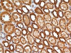
- Experimental details
- Immunohistochemical analysis of MBD1 in human Kidney tissue using MBD1 antibody (Clone: ABM15H2) at 5 µg/ml.
- Protocol
- Protocol
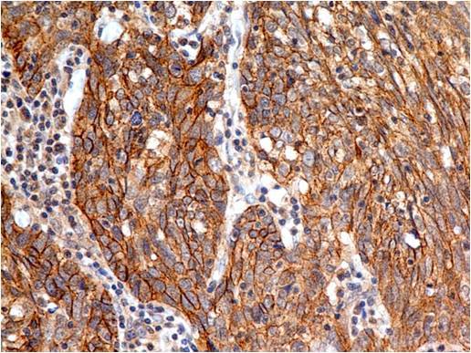
- Submitted by
- ABEOMICS Inc. (provider)
- Main image
- Experimental details
- Immunohistochemical analysis of MBD1 in Squamous cell carcinoma of Lungs using MBD1 antibody (Clone: ABM15H2) at 5 µg/ml.
- Protocol
- Protocol
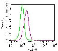
Supportive validation
- Submitted by
- ABEOMICS Inc. (provider)
- Main image
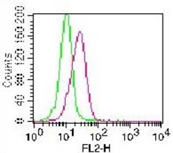
- Experimental details
- Intracellular staining of PMA treated Jurkat cells using 0.5 µg/10^6 Cells. Green represents isotope control, Red represents 10-7007 (ABM15H2) antibody. Goat anti-mouse PE was used as secondary antibody.
- Protocol
- Protocol