Explore
 Validate
 Learn
Learn Immunocytochemistry
Antibody data
- Antibody Data
- Antigen structure
- References [0]
- Comments [0]
- Validations
- Immunocytochemistry [2]
- Chromatin Immunoprecipitation [1]
Submit
Validation data
Reference
Comment
Report error
- Product number
- 712384 - Provider product page

- Provider
- Invitrogen Antibodies
- Product name
- MBD1 Recombinant Polyclonal Antibody (12HCLC)
- Antibody type
- Polyclonal
- Antigen
- Other
- Description
- This antibody is predicted to react with Monkey, Cat, Sheep.
- Antibody clone number
- 12HCLC
- Concentration
- 0.5 mg/mL
No comments: Submit comment
Supportive validation
- Submitted by
- Invitrogen Antibodies (provider)
- Main image
- Experimental details
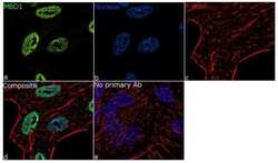
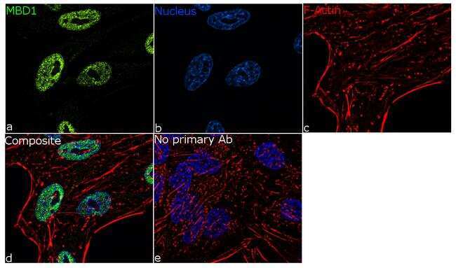
- For immunofluorescence analysis, HeLa cells were fixed and permeabilized for detection of endogenous MBD1 using Anti-MBD1 Recombinant Rabbit Polyclonal Antibody (Product # 712384, 1:100 dilution) and labeled with Goat anti-Rabbit IgG (H+L) Superclonal™ Secondary Antibody, Alexa Fluor® 488 conjugate (Product # A27034, 1:2000). Panel a) shows representative cells that were stained for detection and localization of MBD1 protein (green), Panel b) is stained for nuclei (blue) using ProLong™ Diamond Antifade Mountant with DAPI (Product # P36962). Panel c) represents cytoskeletal F-actin staining using Rhodamine Phalloidin (Product # R415, 1:300). Panel d) is a composite image of Panels a, b and c clearly demonstrating nuclear localization of MBD1. Panel e) represents control cells with no primary antibody to assess background. The images were captured at 60X magnification.
- Submitted by
- Invitrogen Antibodies (provider)
- Main image

- Experimental details
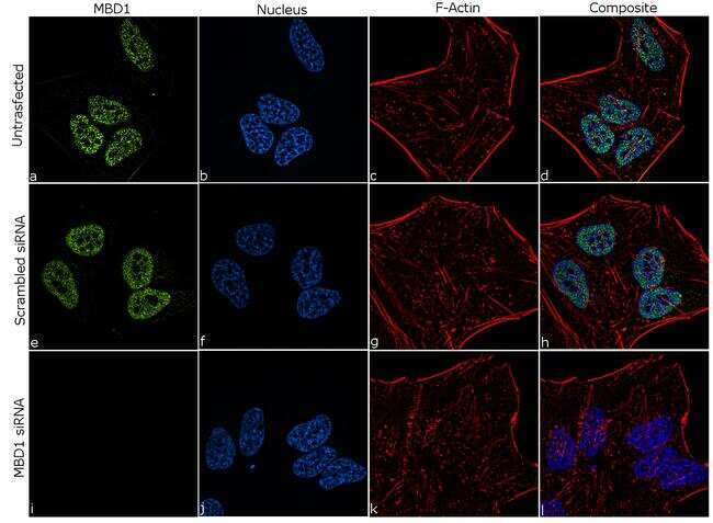
- Knockdown of MBD1 was achieved by transfecting HeLa cells with specific siRNA (Silencer® select Product # s8549, s8547). Immunofluorescence analysis was performed on HeLa cells (untransfected, panel a-d), transfected with MBD1 specific siRNA (panel i-l) or non-specific scrambled siRNA (panels e-h). Cells were fixed, permeabilized, and labeled with Anti-MBD1 Recombinant Rabbit Polyclonal Antibody (Product # 712384, 1:100 dilution), followed by Goat anti-Rabbit IgG (H+L) Superclonal™ Secondary Antibody, Alexa Fluor® 488 conjugate (Product # A27034, 1:2000). Nuclei (blue) were stained using ProLong™ Diamond Antifade Mountant with DAPI (Product # P36962), and Rhodamine Phalloidin (Product # R415, 1:300) was used for cytoskeletal F-actin (red) staining. Significant reduction of signal was observed upon siRNA mediated knockdown (panel i-l) confirming the specificity of the antibody to MBD1 (green). The images were captured at 60X magnification.
Supportive validation
- Submitted by
- Invitrogen Antibodies (provider)
- Main image

- Experimental details
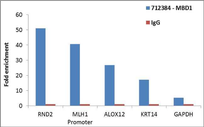
- Enrichment of endogenous MBD1 protein at specific gene loci using Anti-MBD1 Antibody: Chromatin Immunoprecipitation (ChIP) was performed using Anti-MBD1 Recombinant Rabbit Polyclonal Antibody (Product # 712384, 4 µg) on sheared chromatin from 2 million HeLa cells using the MAGnify ChIP System kit (Product # 49-2024). Normal Rabbit IgG was used as a negative IP control. The purified DNA was analyzed by qPCR with PCR primer pairs over RND2, MLH1 promoter, ALOX12, KRT14 (active) and GAPDH gene body (inactive). Data is presented as fold enrichment of the antibody signal versus the negative control IgG using the comparative CT method.