Explore
 Validate
 Learn
Learn Western blot
Antibody data
- Antibody Data
- Antigen structure
- References [3]
- Comments [0]
- Validations
- Western blot [3]
- Immunocytochemistry [1]
Submit
Validation data
Reference
Comment
Report error
- Product number
- MA1-25066 - Provider product page

- Provider
- Invitrogen Antibodies
- Product name
- gamma Adaptin Monoclonal Antibody (100/3)
- Antibody type
- Monoclonal
- Antigen
- Other
- Description
- Recommended positive controls: bovine brain.
- Antibody clone number
- 100/3
- Concentration
- 0.9 mg/mL
Submitted references Intracellular trafficking and activation of the furin proprotein convertase: localization to the TGN and recycling from the cell surface.
Intracellular trafficking and activation of the furin proprotein convertase: localization to the TGN and recycling from the cell surface.
Targeting and mistargeting of plasma membrane adaptors in vitro.
Molloy SS, Thomas L, VanSlyke JK, Stenberg PE, Thomas G
The EMBO journal 1994 Jan 1;13(1):18-33
The EMBO journal 1994 Jan 1;13(1):18-33
Intracellular trafficking and activation of the furin proprotein convertase: localization to the TGN and recycling from the cell surface.
Molloy SS, Thomas L, VanSlyke JK, Stenberg PE, Thomas G
The EMBO journal 1994 Jan 1;13(1):18-33
The EMBO journal 1994 Jan 1;13(1):18-33
Targeting and mistargeting of plasma membrane adaptors in vitro.
Seaman MN, Ball CL, Robinson MS
The Journal of cell biology 1993 Dec;123(5):1093-105
The Journal of cell biology 1993 Dec;123(5):1093-105
No comments: Submit comment
Supportive validation
- Submitted by
- Invitrogen Antibodies (provider)
- Main image
- Experimental details
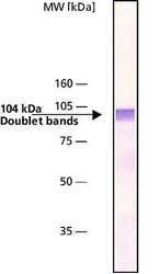
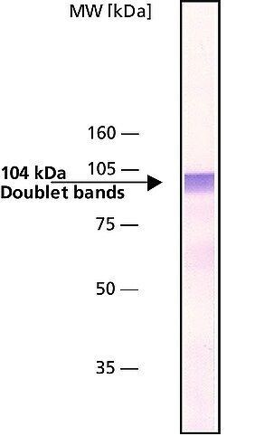
- Western Blot analysis of bovine brain extract using gamma Adaptin Monoclonal Antibody (100/3) (Product # MA1-25066) at 1:200.
- Submitted by
- Invitrogen Antibodies (provider)
- Main image
- Experimental details
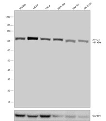
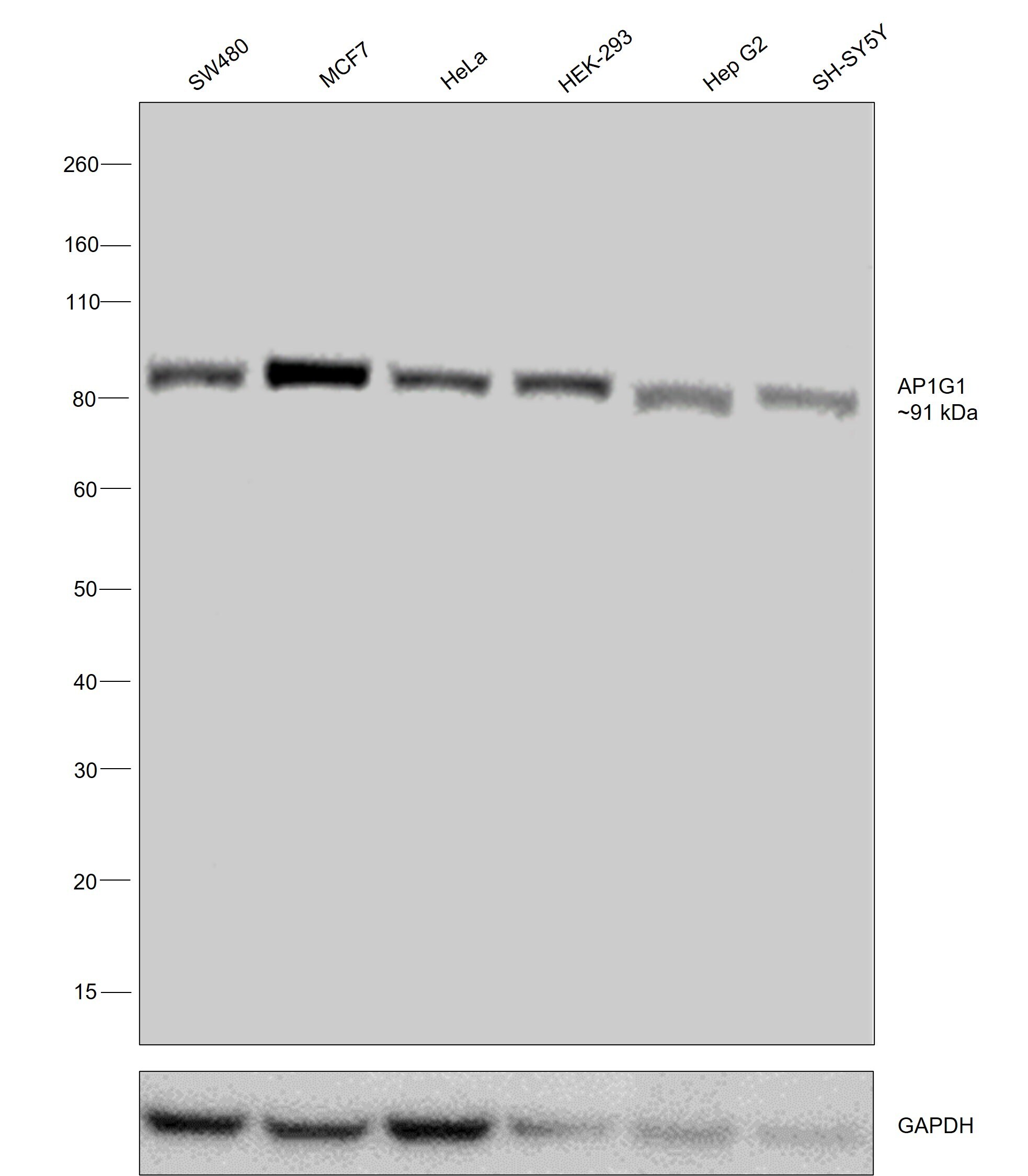
- Western blot was performed using Anti-gamma Adaptin Monoclonal Antibody (100/3) (Product # MA1-25066) and a ~91 kDa band corresponding to AP1G1 was observed across all cell lysates tested. Whole cell extracts (30 µg lysate) of SW480 (Lane 1), MCF7 (Lane 2), HeLa (Lane 3), HEK-293 (Lane 4), Hep G2 (Lane 5), SH-SY5Y (Lane 6) were electrophoresed using NuPAGE™ 4-12% Bis-Tris Protein Gel (Product # NP0322BOX). Resolved proteins were then transferred onto a nitrocellulose membrane (Product # IB23001) by iBlot® 2 Dry Blotting System (Product # IB21001). The blot was probed with the primary antibody (1:200 dilution) and detected by chemiluminescence with Goat anti-Mouse IgG (H+L) Superclonal™ Recombinant Secondary Antibody, HRP (Product # A28177,1:20000 dilution) using the iBright™ FL1500 Imaging System (Product # A44115). Chemiluminescent detection was performed using SuperSignal™ West Pico PLUS Chemiluminescent Substrate (Product # 34580).
- Submitted by
- Invitrogen Antibodies (provider)
- Main image
- Experimental details
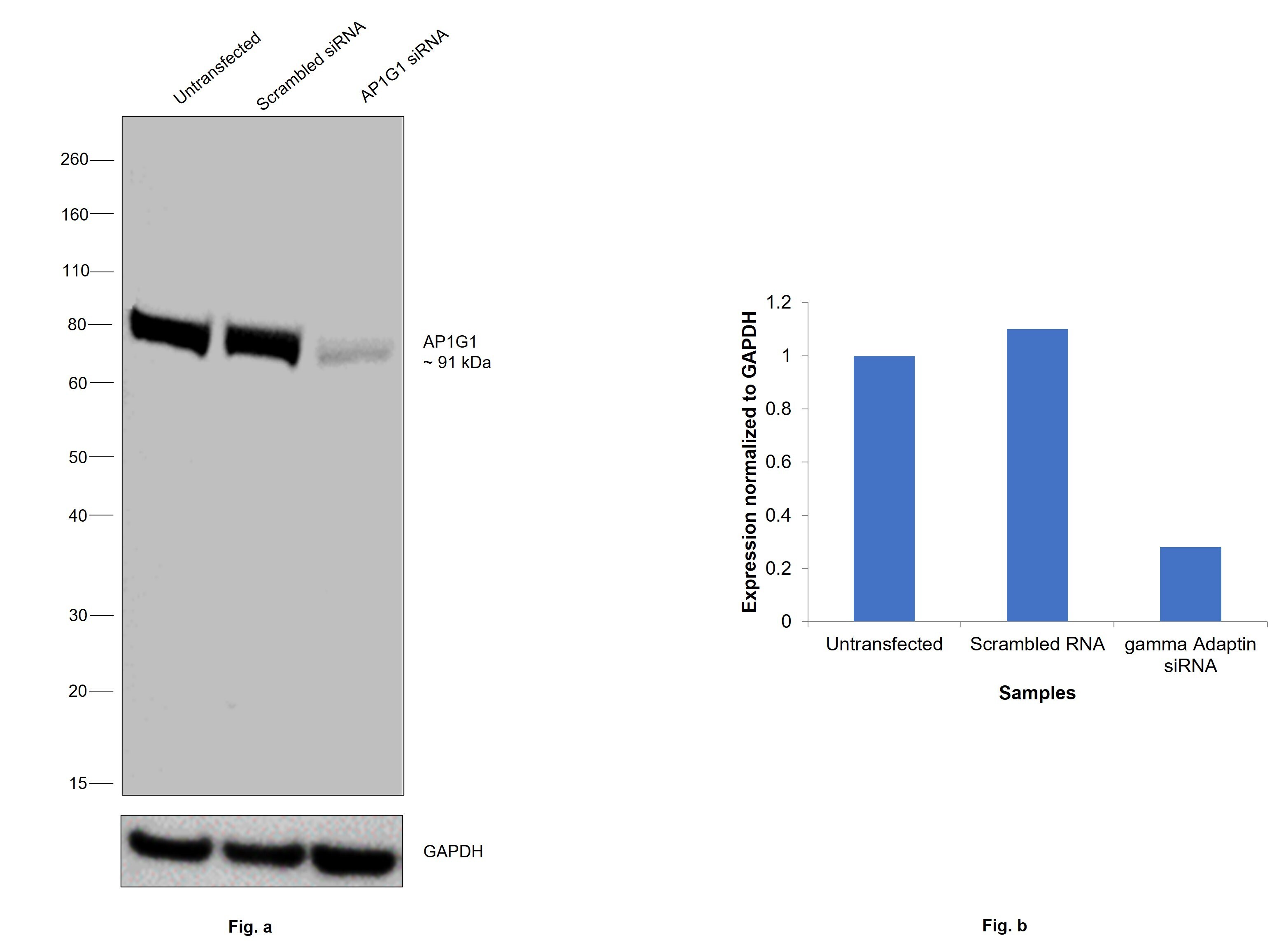
- Knockdown of AP1G1 specific siRNAs (Silencer® select Product # s1142, s1143). Western blot analysis (Fig. a) was performed using Whole cell extracts from the AP1G1knockdown cells (lane 3), non-targeting scrambled siRNA transfected cells (lane 2) and untransfected cells (lane 1). The blot was probed with gamma Adaptin Monoclonal Antibody (100/3) (Product # MA1-25066, 1:200 dilution) and Goat anti-Mouse IgG (H+L) Superclonal™ Recombinant Secondary Antibody, HRP (Product # A28177, 1:20000 dilution). Densitometric analysis of this western blot is shown in histogram (Fig. b). Decrease in signal upon siRNA mediated knock down confirms that antibody is specific to AP1G1.
Supportive validation
- Submitted by
- Invitrogen Antibodies (provider)
- Main image
- Experimental details
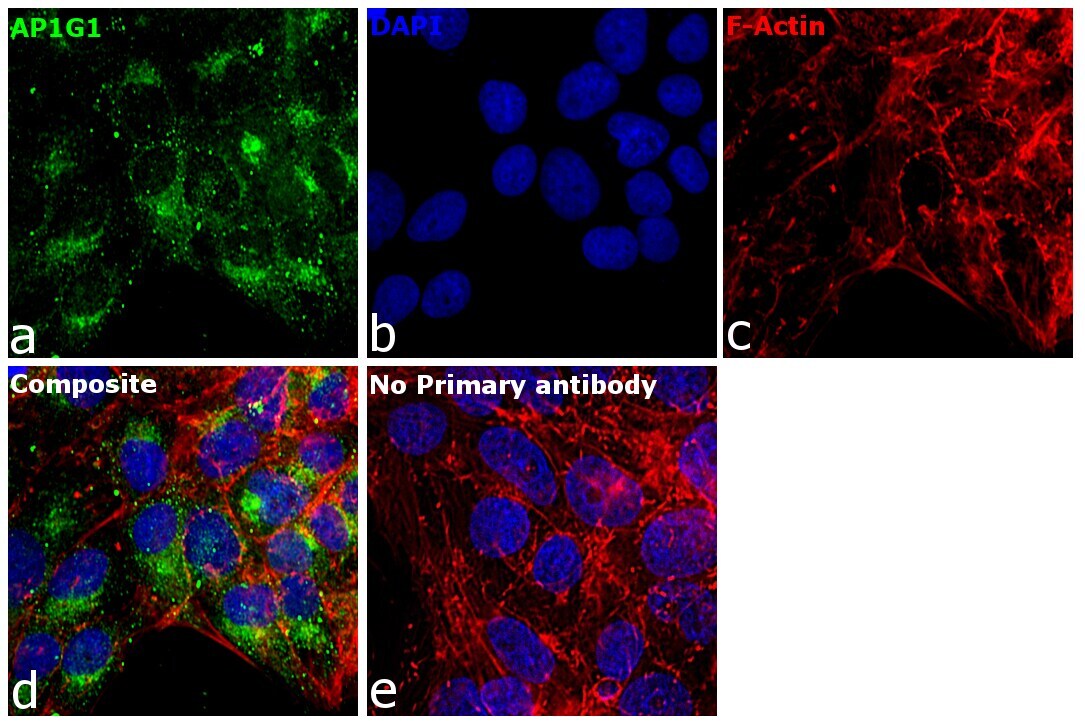
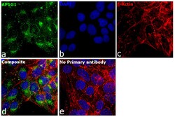
- Immunofluorescence analysis of gamma Adaptin was performed using 70% confluent log phase BeWo cells. The cells were fixed with 4% paraformaldehyde for 10 minutes, permeabilized with 0.1% Triton™ X-100 for 15 minutes, and blocked with 2% BSA for 45 minutes at room temperature. The cells were labeled with gamma Adaptin Monoclonal Antibody (100/3) (Product # MA1-25066) at 1:100 in 0.1% BSA, incubated at 4 degree celsius overnight and then labeled with Donkey anti-Mouse IgG (H+L) Highly Cross-Adsorbed Secondary Antibody, Alexa Fluor Plus 488 (Product # A32766), (1:2000), for 45 minutes at room temperature (Panel a: Green). Nuclei (Panel b:Blue) were stained with Hoechst 33342 (Product # H1399). F-actin (Panel c: Red) was stained with Rhodamine Phalloidin (Product # R415, 1:300). Panel d represents the merged image showing cytoplasmic(Golgi complex like pattern) localization. Panel e represents control cells with no primary antibody to assess background. The images were captured at 40X magnification in CellInsight CX7 LZR High-Content Screening (HCS) Platform (Product # CX7A1110LZR) and externally deconvoluted (D.Sage et al. / Methods 115 (2017) 28–41).