 Explore
 Validate
 Learn
Learn Western blot
Antibody data
- Antibody Data
- Antigen structure
- References [0]
- Comments [0]
- Validations
- Western blot [3]
Submit
Validation data
Reference
Comment
Report error
- Product number
- 44321M - Provider product page

- Provider
- Invitrogen Antibodies
- Product name
- TOP1 Monoclonal Antibody (23B11)
- Antibody type
- Monoclonal
- Antigen
- Synthetic peptide
- Reactivity
- Human
- Host
- Mouse
- Isotype
- IgG
- Antibody clone number
- 23B11
- Vial size
- 50 µL
- Concentration
- 1 mg/mL
- Storage
- -20°C
No comments: Submit comment
Supportive validation
- Submitted by
- Invitrogen Antibodies (provider)
- Main image

- Experimental details
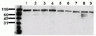
- Whole cell lysates of serum starved HeLa (1), HepG2 (2), HEK293 (3), SH-SY5Y (4), MDCK (5) PC12 (6), CMT 93 (7), Neuro 2A (8) and 3T3 (9) tumor cells (approximately 20,000 cells per lane) were resolved by SDS-PAGE and transferred to PVDF. The membrane was blocked with a casein/Tween 20 buffer, then incubated with mouse anti-Topoisomerase-1 antibody (Product # 44321M) at 0.5 µg/mL for 1 hour at room temperature. After washing, the membrane was incubated with an anti-mouse HRP-conjugated secondary antibody and signals were detected using an ECL detection method (exposure time: 30 seconds).
- Submitted by
- Invitrogen Antibodies (provider)
- Main image

- Experimental details
- Whole cell lysates of serum starved HeLa (1), HepG2 (2), HEK293 (3), SH-SY5Y (4), MDCK (5) PC12 (6), CMT 93 (7), Neuro 2A (8) and 3T3 (9) tumor cells (approximately 20,000 cells per lane) were resolved by SDS-PAGE and transferred to PVDF. The membrane was blocked with a casein/Tween 20 buffer, then incubated with mouse anti-Topoisomerase-1 antibody (Product # 44321M) at 0.5 µg/mL for 1 hour at room temperature. After washing, the membrane was incubated with an anti-mouse HRP-conjugated secondary antibody and signals were detected using an ECL detection method (exposure time: 30 seconds).

- Submitted by
- Invitrogen Antibodies (provider)
- Main image
- Experimental details
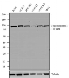
- Western blot analysis was performed on whole cell extracts (30 µg lysate) of Jurkat (Lane 1), MCF-7 (Lane 2), HEK-293 (Lane 3), NIH/3T3 (Lane 4), NTERA-2 (Lane 5) and HeLa (Lane 6). The blots were probed with Anti-Topoisomerase I Mouse Monoclonal Antibody (Product # 44321M, 1:1000 dilution) and detected by chemiluminescence using Goat anti-Mouse IgG (H+L) Superclonal™ Secondary Antibody, HRP conjugate (Product # A28177, 0.4 µg/mL, 1:2500 dilution). A ~ 90 kDa band corresponding to Topoisomerase I was observed across cell lines tested. Known quantity of protein samples were electrophoresed using Novex® NuPAGE® 4-12 % Bis-Tris gel (Product # NP0321BOX), XCell SureLock™ Electrophoresis System (Product # EI0002) and Novex® Sharp Pre-Stained Protein Standard (Product # LC5800). Resolved proteins were then transferred onto a nitrocellulose membrane with Pierce™ Power Blotter System (Product # 22834). The membrane was probed with the relevant primary and secondary Antibody following blocking with 5 % skimmed milk. Chemiluminescent detection was performed using Pierce™ ECL Western Blotting Substrate (Product # 32106).
