 Explore
 Validate
 Learn
Learn Western blot
 Immunocytochemistry
Antibody data
- Antibody Data
- Antigen structure
- References [1]
- Comments [0]
- Validations
- Immunocytochemistry [5]
- Immunohistochemistry [1]
- Flow cytometry [2]
- Chromatin Immunoprecipitation [2]
- Other assay [1]
Submit
Validation data
Reference
Comment
Report error
- Product number
- MA5-32228 - Provider product page

- Provider
- Invitrogen Antibodies
- Product name
- TOP1 Recombinant Rabbit Monoclonal Antibody (SC69-03)
- Antibody type
- Monoclonal
- Antigen
- Synthetic peptide
- Description
- Recombinant rabbit monoclonal antibodies are produced using in vitro expression systems. The expression systems are developed by cloning in the specific antibody DNA sequences from immunoreactive rabbits. Then, individual clones are screened to select the best candidates for production. The advantages of using recombinant rabbit monoclonal antibodies include: better specificity and sensitivity, lot-to-lot consistency, animal origin-free formulations, and broader immunoreactivity to diverse targets due to larger rabbit immune repertoire.
- Reactivity
- Human, Mouse
- Host
- Rabbit
- Isotype
- IgG
- Antibody clone number
- SC69-03
- Vial size
- 100 μL
- Concentration
- 1 mg/mL
- Storage
- Store at 4°C short term. For long term storage, store at -20°C, avoiding freeze/thaw cycles.
Submitted references A brain proteomic signature of incipient Alzheimer's disease in young APOE ε4 carriers identifies novel drug targets.
Roberts JA, Varma VR, An Y, Varma S, Candia J, Fantoni G, Tiwari V, Anerillas C, Williamson A, Saito A, Loeffler T, Schilcher I, Moaddel R, Khadeer M, Lovett J, Tanaka T, Pletnikova O, Troncoso JC, Bennett DA, Albert MS, Yu K, Niu M, Haroutunian V, Zhang B, Peng J, Croteau DL, Resnick SM, Gorospe M, Bohr VA, Ferrucci L, Thambisetty M
Science advances 2021 Nov 12;7(46):eabi8178
Science advances 2021 Nov 12;7(46):eabi8178
No comments: Submit comment
Supportive validation
- Submitted by
- Invitrogen Antibodies (provider)
- Main image

- Experimental details
- Immunocytochemical analysis of Topoisomerase I in Hela cells using a Topoisomerase I Monoclonal antibody (Product # MA5-32228) as seen in green. The nuclear counter stain is DAPI (blue). Cells were fixed in paraformaldehyde, permeabilised with 0.25% Triton X100/PBS.
- Submitted by
- Invitrogen Antibodies (provider)
- Main image

- Experimental details
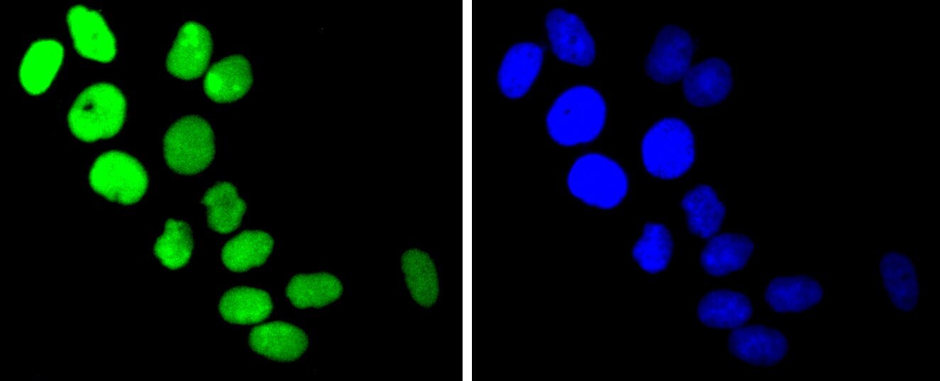
- Immunocytochemical analysis of Topoisomerase I in SHG-44 cells using a Topoisomerase I Monoclonal antibody (Product # MA5-32228) as seen in green. The nuclear counter stain is DAPI (blue). Cells were fixed in paraformaldehyde, permeabilised with 0.25% Triton X100/PBS.

- Submitted by
- Invitrogen Antibodies (provider)
- Main image
- Experimental details
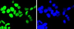
- Immunocytochemical analysis of Topoisomerase I in 293 cells using a Topoisomerase I Monoclonal antibody (Product # MA5-32228) as seen in green. The nuclear counter stain is DAPI (blue). Cells were fixed in paraformaldehyde, permeabilised with 0.25% Triton X100/PBS.

- Submitted by
- Invitrogen Antibodies (provider)
- Main image

- Experimental details
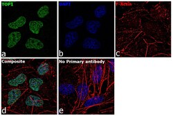
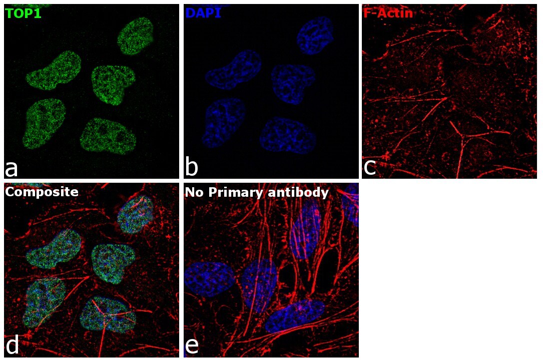
- Immunofluorescence analysis of TOP1 was performed using 70% confluent log phase HeLa cells. The cells were fixed with 4% paraformaldehyde for 10 minutes, permeabilized with 0.1% Triton™ X-100 for 15 minutes, and blocked with 2% BSA for 1 hour at room temperature. The cells were labeled with TOP1 Recombinant Rabbit Monoclonal Antibody (SC69-03) (Product # MA5-32228) at 5 µg/mL in 0.1% BSA, incubated at 4 degree Celsius overnight and then labeled with Goat anti-Rabbit IgG (H+L), Superclonal™ Recombinant Secondary Antibody, Alexa Fluor 488 (Product # A27034) at a dilution of 1:2000 for 45 minutes at room temperature (Panel a: green). Nuclei (Panel b: blue) were stained with ProLong™ Diamond Antifade Mountant with DAPI (Product # P36962). F-actin (Panel c: red) was stained with Rhodamine Phalloidin (Product # R415). Panel d represents the merged image showing nuclear localization. Panel e represents control cells with no primary antibody to assess background. The images were captured at 60X magnification.

- Submitted by
- Invitrogen Antibodies (provider)
- Main image
- Experimental details
- Immunofluorescence analysis of TOP1 was performed using 70% confluent log phase HeLa cells. The cells were fixed with 4% paraformaldehyde for 10 minutes, permeabilized with 0.1% Triton™ X-100 for 15 minutes, and blocked with 2% BSA for 1 hour at room temperature. The cells were labeled with TOP1 Recombinant Rabbit Monoclonal Antibody (SC69-03) (Product # MA5-32228) at 5 µg/mL in 0.1% BSA, incubated at 4 degree Celsius overnight and then labeled with Goat anti-Rabbit IgG (Heavy Chain), Superclonal™ Recombinant Secondary Antibody, Alexa Fluor 488 (Product # A27034) at a dilution of 1:2000 for 45 minutes at room temperature (Panel a: green). Nuclei (Panel b: blue) were stained with ProLong™ Diamond Antifade Mountant with DAPI (Product # P36962). F-actin (Panel c: red) was stained with Rhodamine Phalloidin (Product # R415). Panel d represents the merged image showing nuclear localization. Panel e represents control cells with no primary antibody to assess background. The images were captured at 60X magnification.
Supportive validation
- Submitted by
- Invitrogen Antibodies (provider)
- Main image
- Experimental details
- Immunohistochemical analysis of Topoisomerase I of paraffin-embedded Mouse brain tissue using a Topoisomerase-I Monoclonal antibody (Product #MA5-32228). Counter stained with hematoxylin.
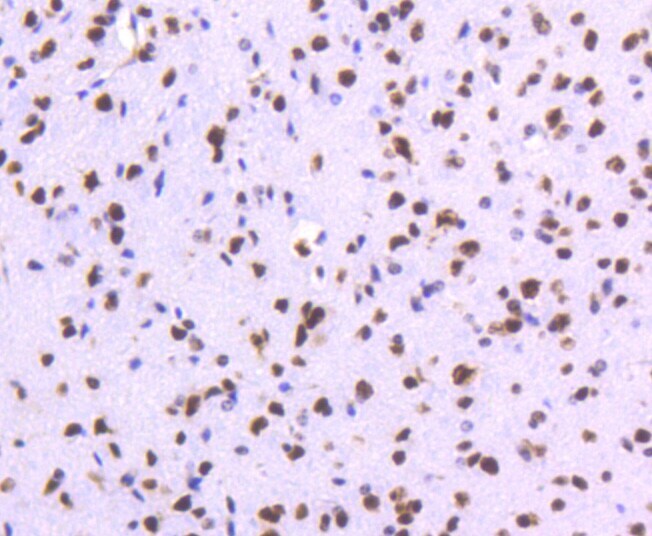
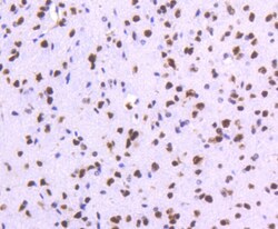
Supportive validation
- Submitted by
- Invitrogen Antibodies (provider)
- Main image
- Experimental details
- Flow Cytometric analysis of Topoisomerase I in HepG2 cells using a Topoisomerase I Monoclonal Antibody (Product # MA5-32228) at a dilution of 1:50, as seen in red compared with an unlabelled control (cells without incubation with primary antibody; black). Alexa Fluor 488-conjugated goat anti rabbit IgG was used as the secondary antibody.
- Submitted by
- Invitrogen Antibodies (provider)
- Main image

- Experimental details
- Flow Cytometric analysis of Topoisomerase I in HepG2 cells using a Topoisomerase I Monoclonal Antibody (Product # MA5-32228) at a dilution of 1:50, as seen in red compared with an unlabelled control (cells without incubation with primary antibody; black). Alexa Fluor 488-conjugated goat anti rabbit IgG was used as the secondary antibody.

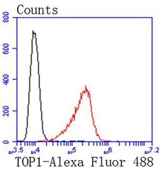
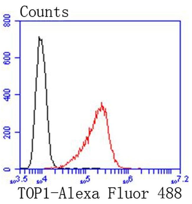
Supportive validation
- Submitted by
- Invitrogen Antibodies (provider)
- Main image

- Experimental details
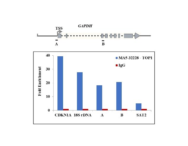
- Chromatin Immunoprecipitation (ChIP) assay of endogenous TOP1 protein using Anti-TOP1 Antibody: ChIP was performed using Anti-TOP1 Rabbit Recombinant Monoclonal Antibody (Product # MA5-32228, 5 µg) on sheared chromatin from Camptothecin treated HCT 116 cells (10uM, 2hours) (using the MAGnify ChIP System kit (Product # 49-2024). Normal Rabbit IgG was used as a negative IP control. The purified DNA was analyzed by qPCR using primers binding to CDKN1A intron 1, 18s rDNA, promoter and transcriptional start site of GAPDH, and SAT2 satellite repeats. Data is presented as fold enrichment of the antibody signal versus the negative control IgG using the comparative CT method.
- Submitted by
- Invitrogen Antibodies (provider)
- Main image

- Experimental details
- Chromatin Immunoprecipitation (ChIP) assay of endogenous TOP1 protein using Anti-TOP1 Antibody: ChIP was performed using Anti-TOP1 Rabbit Recombinant Monoclonal Antibody (Product # MA5-32228, 5 µg) on sheared chromatin from Camptothecin treated HCT 116 cells (10uM, 2hours) (using the MAGnify ChIP System kit (Product # 49-2024). Normal Rabbit IgG was used as a negative IP control. The purified DNA was analyzed by qPCR using primers binding to CDKN1A intron 1, 18s rDNA, promoter and transcriptional start site of GAPDH, and SAT2 satellite repeats. Data is presented as fold enrichment of the antibody signal versus the negative control IgG using the comparative CT method.

Supportive validation
- Submitted by
- Invitrogen Antibodies (provider)
- Main image
- Experimental details
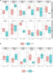
- Fig. 5. Primary validation of incipient AD proteomic signature. Results of primary validation analyses performed using Western blotting in 3xTg-AD transgenic mice (AD) compared to wild type (WT) ( A ) and human brain tissue samples comparing AD to CN ( B ). The 3xTg-AD ( n = 6) and WT ( n = 6) mice included young (38 to 40 weeks) and old (83 to 136 weeks) mice. (A) Box plots represent differences in protein levels in brain homogenates between 3xTg-AD (blue) and WT (red) mice. The top and bottom rows indicate protein levels in young and old mice, respectively. (B) Box plots represent differences in protein levels in brain homogenates between AD (blue; n = 11) and CN (red; n = 11). Protein levels that were significantly different between 3xTg-AD and WT or AD and CN are indicated with * ( P < 0.05) or ** ( P < 0.01). 3xTg-AD, 3xTg-AD transgenic mouse model of AD; AD, Alzheimer's disease; WT, wild type; DUSP3, dual-specificity phosphate 3; STAT3, signal transducer and activator of transcription 3; TOP1, DNA topoisomerase I; FYN1, FYN proto-oncogene tyrosine kinase; LGALS8, galectin-8; YES1, YES proto-oncogene tyrosine kinase.
