Explore
 Validate
 Learn
Learn Western blot
 Immunocytochemistry
Antibody data
- Antibody Data
- Antigen structure
- References [1]
- Comments [0]
- Validations
- Immunocytochemistry [1]
- Immunohistochemistry [1]
Submit
Validation data
Reference
Comment
Report error
- Product number
- HPA019039 - Provider product page

- Provider
- Atlas Antibodies
- Proper citation
- Atlas Antibodies Cat#HPA019039, RRID:AB_1858187
- Product name
- Anti-TOP1
- Antibody type
- Polyclonal
- Description
- Polyclonal Antibody against Human TOP1, Gene description: topoisomerase (DNA) I, Validated applications: WB, IHC, ICC, Uniprot ID: P11387, Storage: Store at +4°C for short term storage. Long time storage is recommended at -20°C.
- Reactivity
- Human, Mouse, Rat
- Host
- Rabbit
- Conjugate
- Unconjugated
- Isotype
- IgG
- Vial size
- 100 µl
- Concentration
- 0.2 mg/ml
- Storage
- Store at +4°C for short term storage. Long time storage is recommended at -20°C.
- Handling
- The antibody solution should be gently mixed before use.
Submitted references An integrated genome-wide multi-omics analysis of gene expression dynamics in the preimplantation mouse embryo
Israel S, Ernst M, Psathaki O, Drexler H, Casser E, Suzuki Y, Makalowski W, Boiani M, Fuellen G, Taher L
Scientific Reports 2019;9(1)
Scientific Reports 2019;9(1)
No comments: Submit comment
Supportive validation
- Submitted by
- Atlas Antibodies (provider)
- Main image
- Experimental details
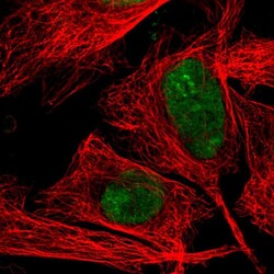
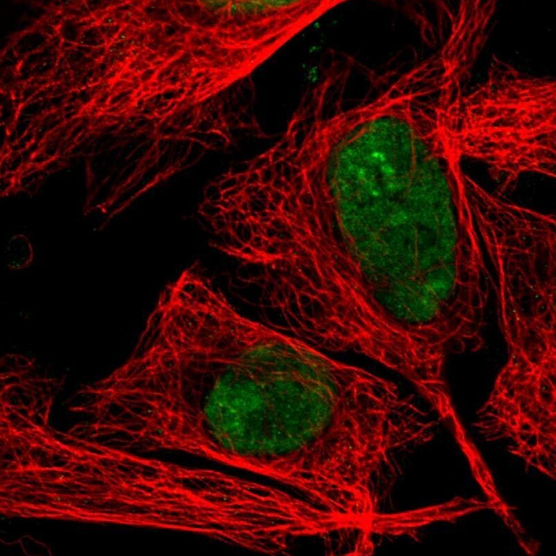
- Immunofluorescent staining of human cell line U-2 OS shows localization to nucleus & nucleoli fibrillar center.
- Sample type
- Human
Supportive validation
- Submitted by
- Atlas Antibodies (provider)
- Enhanced method
- Orthogonal validation
- Main image
- Experimental details
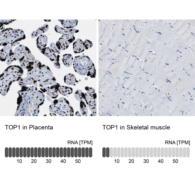
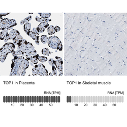
- Immunohistochemistry analysis in human placenta and skeletal muscle tissues using HPA019039 antibody. Corresponding TOP1 RNA-seq data are presented for the same tissues.
- Sample type
- Human
- Protocol
- Protocol