Explore
 Validate
 Learn
Learn Western blot
Antibody data
- Antibody Data
- Antigen structure
- References [0]
- Comments [0]
- Validations
- Western blot [6]
- Immunocytochemistry [1]
- Immunohistochemistry [2]
- Flow cytometry [1]
Submit
Validation data
Reference
Comment
Report error
- Product number
- MA5-17129 - Provider product page

- Provider
- Invitrogen Antibodies
- Product name
- MSH6 Monoclonal Antibody (3E1)
- Antibody type
- Monoclonal
- Antigen
- Purifed from natural sources
- Description
- MA5-17129 targets MSH6 in FACS, ICC, IHC, pep-ELISA, and WB applications and shows reactivity with Human samples.
- Antibody clone number
- 3E1
- Concentration
- Conc. Not Determined
No comments: Submit comment
Supportive validation
- Submitted by
- Invitrogen Antibodies (provider)
- Main image

- Experimental details
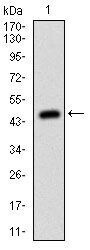
- Western blot analysis of MSH6 using a MSH6 monoclonal antibody (Product # MA5-17129) against a human MSH6 (AA: 217-395) recombinant protein.

- Submitted by
- Invitrogen Antibodies (provider)
- Main image
- Experimental details
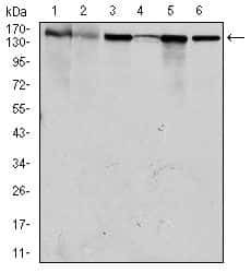
- Western blot analysis of MSH6 using MSH6 monoclonal antibody (Product # MA5-17129) in HEK293 (1), HCT116 (2), A549 (3), A431 (4), MCF-7 (5) and HepG2 (6) cell lysate.

- Submitted by
- Invitrogen Antibodies (provider)
- Main image

- Experimental details
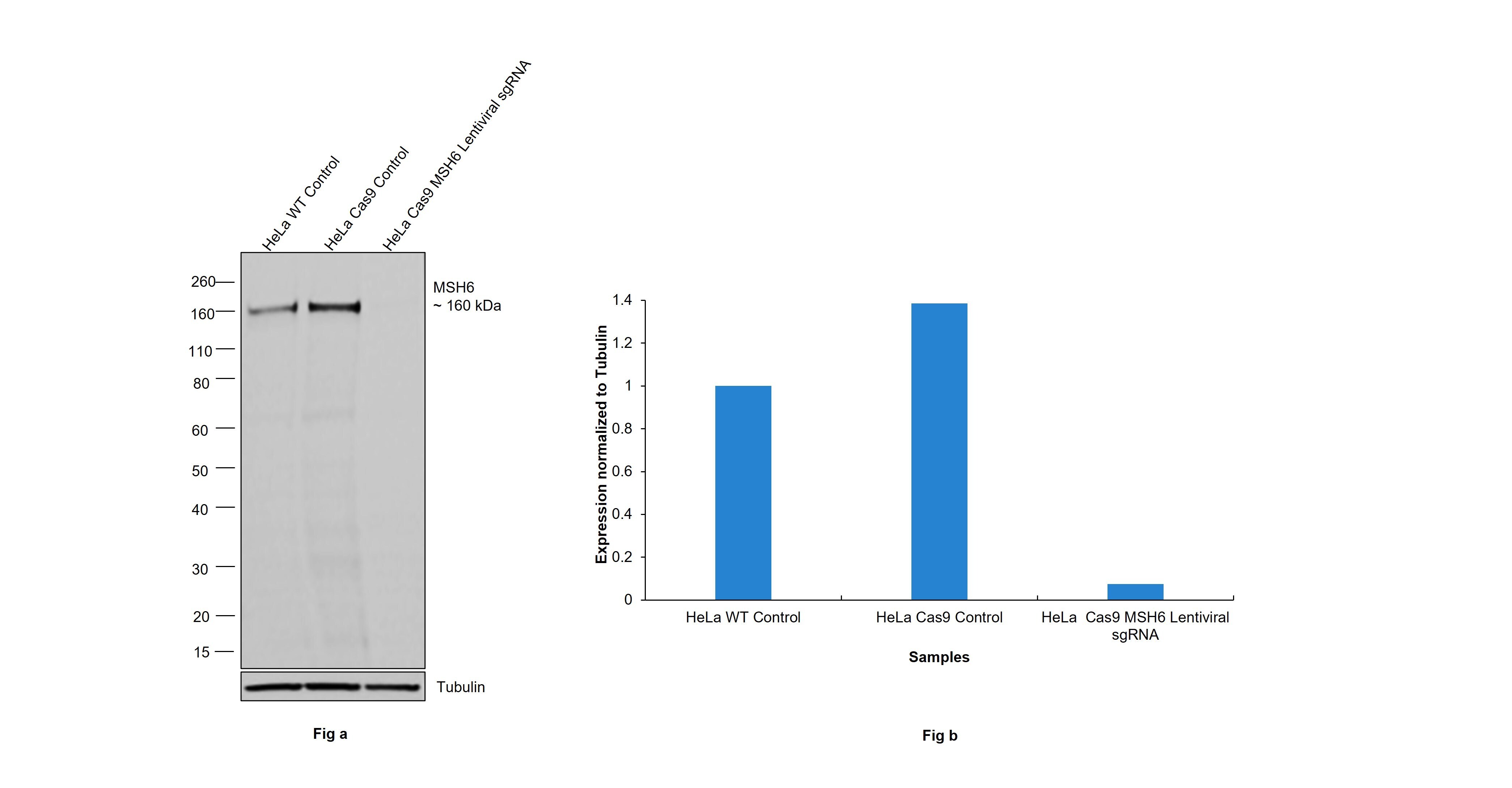
- CRISPR-Cas9 mediated genome editing ofMSH6 (as confirmed by next generation sequencing) was achieved by using LentiArray™ Lentiviral sgRNA (Product # A32042, Assay ID CRISPR1065222_LV) and LentiArray Cas9 Lentivirus (Product # A32064). Fig (a) Western blot analysis of MSH6 was performed by loading 30 µg of HeLa wild type (Lane 1), HeLa Cas9 (Lane 2) and HeLa Cas9 cells transduced with MSH6 Lentiviral sgRNA (Lane 3) modified whole cell extracts. The samples were electrophoresed using NuPAGE™ 3-8% Tris-Acetate Protein Gel (Product # EA0378BOX). Resolved proteins were then transferred onto a nitrocellulose membrane (Product # IB23001) by iBlot® 2 Dry Blotting System (Product # IB21001). The blot was probed with Anti-MSH6 Monoclonal Antibody (3E1) (Product # MA5-17129) using 1:1000 dilution and Goat anti-Mouse IgG (H+L) Superclonal™ Recombinant Secondary Antibody, HRP (Product # A28177 1:5000 dilution).Chemiluminescent detection was performed using Novex® ECL Chemiluminescent Substrate Reagent Kit (Product # WP20005). A loss of signal in sgRNA transduced cells using the LentiArray™ CRISPR product line confirms that antibody is specific toMSH6 (Fig (b)).
- Submitted by
- Invitrogen Antibodies (provider)
- Main image
- Experimental details
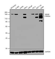
- Western blot was performed using Anti-MSH6 Monoclonal Antibody (3E1) (Product # MA5-17129) and a 160kDa band corresponding to MSH6 was observed in all the tested cell models, except Jurkat and LNCaP which are reported to be negative. This target undergoes heavy phosphorylation and multiple bands between 120-160kDa can be expected. Modified whole cell lysate (1% SDS) (30ug lysate) of SH-SY5Y (Lane 1), K-562 (Lane 2), Jurkat (Lane 3), HeLa (Lane 4), LNCaP (Lane 5), PC-3 (Lane 6), HCT 116 (Lane 7) and A549 (Lane 8) were electrophoresed using NuPAGE® 10 % Bis-Tris gel (Product # NP0302BOX). Resolved proteins were then transferred onto a nitrocellulose membrane (Product # IB23001) by iBlot® 2 Dry Blotting System (Product # IB21001). The blot was probed with the primary antibody (1:1000 dilution) and detected by chemiluminescence with Goat anti-Mouse IgG (H+L) Superclonal™ Recombinant Secondary Antibody, HRP (Product # A28177, 1:4000 dilution) using the iBright FL 1000 (Product # A32752). Chemiluminescent detection was performed using Novex® ECL Chemiluminescent Substrate Reagent Kit (Product # WP20005).

- Submitted by
- Invitrogen Antibodies (provider)
- Main image
- Experimental details
- Western blot analysis of MSH6 using a MSH6 monoclonal antibody (Product # MA5-17129) against a human MSH6 (AA: 217-395) recombinant protein.

- Submitted by
- Invitrogen Antibodies (provider)
- Main image

- Experimental details
- Western blot analysis of MSH6 using MSH6 monoclonal antibody (Product # MA5-17129) in HEK293 (1), HCT116 (2), A549 (3), A431 (4), MCF-7 (5) and HepG2 (6) cell lysate.

Supportive validation
- Submitted by
- Invitrogen Antibodies (provider)
- Main image

- Experimental details
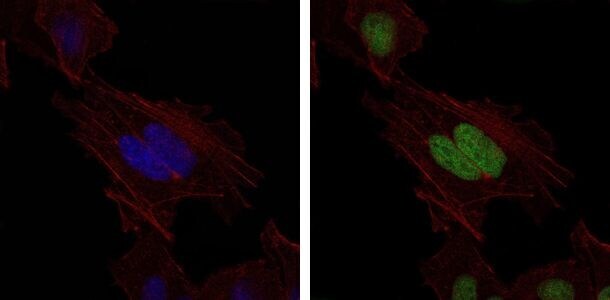
- Immunofluorescence analysis of HeLa cells using MSH6 monoclonal antibody (Product # MA5-17129) (Green). Blue: DRAQ5 fluorescent DNA dye. Red: actin filaments have been labeled with phalloidin.
Supportive validation
- Submitted by
- Invitrogen Antibodies (provider)
- Main image

- Experimental details
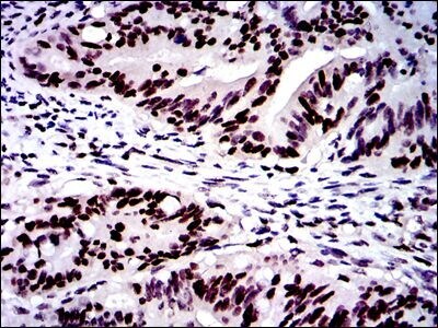
- Immunohistochemical analysis of paraffin-embedded rectum cancer tissues using MSH6 monoclonal antibody (Product # MA5-17129) followed with DAB staining.
- Submitted by
- Invitrogen Antibodies (provider)
- Main image

- Experimental details
- Immunohistochemical analysis of paraffin-embedded colon cancer tissues using MSH6 monoclonal antibody (Product # MA5-17129) followed with DAB staining.

Supportive validation
- Submitted by
- Invitrogen Antibodies (provider)
- Main image

- Experimental details
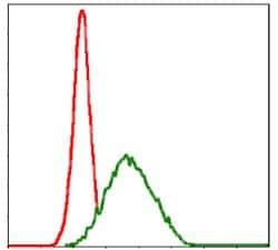
- Flow cytometric analysis of MCF-7 cells using MSH6 monoclonal antibody (Product # MA5-17129) (green) and negative control (red).