 Explore
 Validate
 Learn
Learn Western blot
 Immunoelectron microscopy
Antibody data
- Antibody Data
- Antigen structure
- References [3]
- Comments [0]
- Validations
- Western blot [4]
- Immunocytochemistry [1]
- Immunoprecipitation [1]
- Immunohistochemistry [1]
Submit
Validation data
Reference
Comment
Report error
- Product number
- GTX111661 - Provider product page

- Provider
- GeneTex
- Proper citation
- GeneTex Cat#GTX111661, RRID:AB_1950949
- Product name
- MSH6 antibody [C1C2], Internal
- Antibody type
- Polyclonal
- Reactivity
- Human, Mouse, Zebrafish
- Host
- Rabbit
Submitted references Excluding Lynch syndrome in a female patient with metachronous DNA mismatch repair deficient colon- and ovarian cancer.
GLI1 interferes with the DNA mismatch repair system in pancreatic cancer through BHLHE41-mediated suppression of MLH1.
Sublethal levels of cadmium down-regulate the gene expression of DNA mismatch recognition protein MutS homolog 6 (MSH6) in zebrafish (Danio rerio) embryos.
Crobach S, Jansen AML, Ligtenberg MJL, Koopmans M, Nielsen M, Hes FJ, Wijnen JT, Dinjens WNM, van Wezel T, Morreau H
Familial cancer 2018 Jul;17(3):415-420
Familial cancer 2018 Jul;17(3):415-420
GLI1 interferes with the DNA mismatch repair system in pancreatic cancer through BHLHE41-mediated suppression of MLH1.
Inaguma S, Riku M, Hashimoto M, Murakami H, Saga S, Ikeda H, Kasai K
Cancer research 2013 Dec 15;73(24):7313-23
Cancer research 2013 Dec 15;73(24):7313-23
Sublethal levels of cadmium down-regulate the gene expression of DNA mismatch recognition protein MutS homolog 6 (MSH6) in zebrafish (Danio rerio) embryos.
Hsu T, Tsai HT, Huang KM, Luan MC, Hsieh CR
Chemosphere 2010 Oct;81(6):748-54
Chemosphere 2010 Oct;81(6):748-54
No comments: Submit comment
Supportive validation
- Submitted by
- GeneTex (provider)
- Main image
- Experimental details
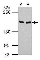
- MSH6 antibody [C1C2], Internal detects MSH6 protein by Western blot analysis. A. 30 ?g Neuro2A whole cell lysate/extract B. 30 ?g GL261 whole cell lysate/extract 5% SDS-PAGE MSH6 antibody [C1C2], Internal (GTX111661) dilution: 1:1000
- Submitted by
- GeneTex (provider)
- Main image
- Experimental details
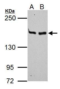
- Sample (30 ug of whole cell lysate) A: Hep G2 (GTX27900) 5% SDS PAGE GTX111661 diluted at 1:1000

- Submitted by
- GeneTex (provider)
- Main image
- Experimental details
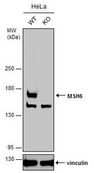
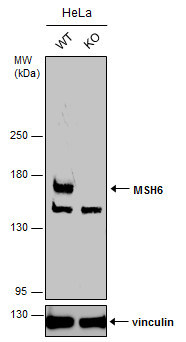
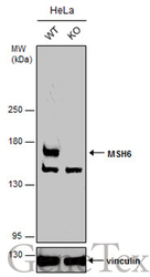
- Wild-type (WT) and MSH6 knockout (KO) HeLa cell extracts (30 ?g) were separated by 5% SDS-PAGE, and the membrane was blotted with MSH6 antibody [C1C2], Internal (GTX111661) diluted at 1:1000. The HRP-conjugated anti-rabbit IgG antibody (GTX213110-01) was used to detect the primary antibody.
- Submitted by
- GeneTex (provider)
- Main image
- Experimental details
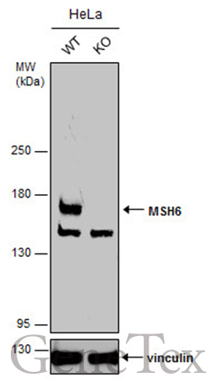
- Wild-type (WT) and MSH6 knockout (KO) HeLa cell extracts (30 ?g) were separated by 5% SDS-PAGE, and the membrane was blotted with MSH6 antibody [C1C2], Internal (GTX111661) diluted at 1:1000. The HRP-conjugated anti-rabbit IgG antibody (GTX213110-01) was used to detect the primary antibody.
Supportive validation
- Submitted by
- GeneTex (provider)
- Main image
- Experimental details
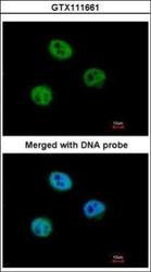
- Immunofluorescence analysis of paraformaldehyde-fixed HeLa, using GTBP (MSH6)(GTX111661) antibody at 1:500 dilution.

Supportive validation
- Submitted by
- GeneTex (provider)
- Main image
- Experimental details
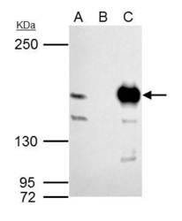
- MSH6 antibody [C1C2], Internal immunoprecipitates MSH6 protein in IP experiments.IP samples: 293T whole cell extractA. 40 £gg 293T whole cell extractB. Control with 4 £gg of preimmune Rabbit IgGC. Immunoprecipitation of MSH6 protein by 4 £gg MSH6 antibody [C1C2], Internal (GTX111661)5 % SDS-PAGEThe immunoprecipitated MSH6 protein was detected by MSH6 antibody [C1C2], Internal (GTX111661) diluted at 1:1000.[EasyBlot anti-rabbit IgG (GTX221666-01) was used as a secondary reagent]

Supportive validation
- Submitted by
- GeneTex (provider)
- Main image
- Experimental details
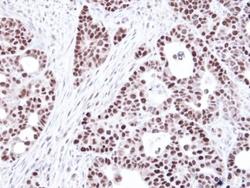
- Immunohistochemical analysis of paraffin-embedded NCIN87 Xenograft, using GTBP (MSH6)(GTX111661) antibody at 1:100 dilution.
