 Explore
 Validate
 Learn
Learn Western blot
 Immunocytochemistry
 Gel shift
Antibody data
- Antibody Data
- Antigen structure
- References [1]
- Comments [0]
- Validations
- Immunocytochemistry [1]
- Immunoprecipitation [1]
- Immunohistochemistry [1]
- Other assay [2]
Submit
Validation data
Reference
Comment
Report error
- Product number
- PA5-29348 - Provider product page

- Provider
- Invitrogen Antibodies
- Product name
- MSH6 Polyclonal Antibody
- Antibody type
- Polyclonal
- Antigen
- Recombinant full-length protein
- Description
- Recommended positive controls: HeLa, HepG2, Neuro2A, GL261. Predicted reactivity: Mouse (88%), Rhesus Monkey (99%), Bovine (93%). Store product as a concentrated solution. Centrifuge briefly prior to opening the vial.
- Reactivity
- Human, Mouse, Zebrafish
- Host
- Rabbit
- Isotype
- IgG
- Vial size
- 100 μL
- Concentration
- 1 mg/mL
- Storage
- Store at 4°C short term. For long term storage, store at -20°C, avoiding freeze/thaw cycles.
Submitted references Establishment of three novel cell lines derived from African American patients with colorectal carcinoma: A unique tool for assessing racial health disparity.
Paredes J, Ji P, Lacomb JF, Shroyer KR, Martello LA, Williams JL
International journal of oncology 2018 Oct;53(4):1516-1528
International journal of oncology 2018 Oct;53(4):1516-1528
No comments: Submit comment
Supportive validation
- Submitted by
- Invitrogen Antibodies (provider)
- Main image
- Experimental details
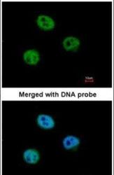
- Immunofluorescent analysis of MSH6 in paraformaldehyde-fixed HeLa cells using a MSH6 polyclonal antibody (Product # PA5-29348) at a 1:500 dilution.

Supportive validation
- Submitted by
- Invitrogen Antibodies (provider)
- Main image

- Experimental details
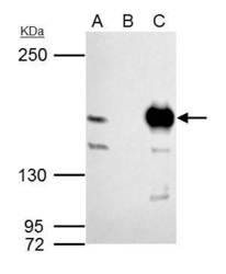
- MSH6 Polyclonal Antibody immunoprecipitates MSH6 protein in IP experiments. IP samples: 293T whole cell extract. A. 40 µg 293T whole cell extract. B. Control with 4 µg of preimmune Rabbit IgG. C. Immunoprecipitation of MSH6 protein by 4 µg MSH6 Polyclonal Antibody (Product # PA5-29348). 5 % SDS-PAGE. The immunoprecipitated MSH6 protein was detected by MSH6 Polyclonal Antibody (Product # PA5-29348) diluted at 1:1,000.

Supportive validation
- Submitted by
- Invitrogen Antibodies (provider)
- Main image
- Experimental details
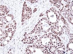
- Immunohistochemical analysis of paraffin-embedded NCIN87 Xenograft, using GTBP (MSH6) (Product # PA5-29348) antibody at 1:100 dilution. Antigen Retrieval: EDTA based buffer, pH 8.0, 15 min.

Supportive validation
- Submitted by
- Invitrogen Antibodies (provider)
- Main image
- Experimental details
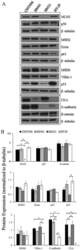
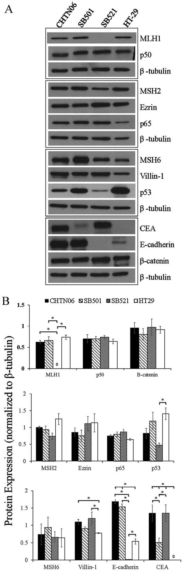
- Figure 3 The expression of proteins associated with colorectal carcinoma (CRC) tumorigenesis and metastasis was determined in the novel African American CRC lines by immunoblotting. (A) Qualitative analysis of CHTN06, SB501 and SB521 and HT-29, a Caucasian American CRC cell line, for protein expression of beta-catenin, p53, nuclear factor (NF)-kappaB (p50 and p65), villin-1, MSH2, MSH6, MLH1 and ezrin. (B) Semi-quantitative densitometry was performed by normalizing protein expression to the respective beta-tubulin loading control. Data were generated from three independent experiments. CEA, carcinoembryonic antigen.
- Submitted by
- Invitrogen Antibodies (provider)
- Main image
- Experimental details
- MSH6 Polyclonal Antibody immunoprecipitates MSH6 protein in IP experiments. IP samples: 293T whole cell extract. A. 40 µg 293T whole cell extract. B. Control with 4 µg of preimmune Rabbit IgG. C. Immunoprecipitation of MSH6 protein by 4 µg MSH6 Polyclonal Antibody (Product # PA5-29348). 5 % SDS-PAGE. The immunoprecipitated MSH6 protein was detected by MSH6 Polyclonal Antibody (Product # PA5-29348) diluted at 1:1,000.
