Explore
 Validate
 Learn
Learn Western blot
Antibody data
- Antibody Data
- Antigen structure
- References [1]
- Comments [0]
- Validations
- Western blot [5]
- Immunocytochemistry [2]
- Immunohistochemistry [2]
- Other assay [1]
Submit
Validation data
Reference
Comment
Report error
- Product number
- PA5-19918 - Provider product page

- Provider
- Invitrogen Antibodies
- Product name
- MyD88 Polyclonal Antibody
- Antibody type
- Polyclonal
- Antigen
- Synthetic peptide
- Description
- A suggested positive control is Jurkat cell lysate. PA5-19918 can be used with blocking peptide PEP-0044. The PA5-19918 The immunogen is located within amino acids 220 - 270 of MYD88. Predicted molecular ~ 35kD. In Western blot applications, this antibody has been observed to detect a band at: 35kD Predicted species reactivity based on immunogen sequence: Pig: (100%), Sheep: (100%), Bovine: (100%), Chicken: (100%), Rat: (88%), Mouse (88%)
- Reactivity
- Human, Mouse, Rat
- Host
- Rabbit
- Isotype
- IgG
- Vial size
- 100 µg
- Concentration
- 1.0 mg/mL
- Storage
- Maintain refrigerated at 2-8°C for up to 3 months. For long term storage store at -20°C
Submitted references Cerebral endothelial cell derived small extracellular vesicles improve cognitive function in aged diabetic rats.
Zhang L, Li C, Huang R, Teng H, Zhang Y, Zhou M, Liu X, Fan B, Luo H, He A, Zhao A, Lu M, Chopp M, Zhang ZG
Frontiers in aging neuroscience 2022;14:926485
Frontiers in aging neuroscience 2022;14:926485
No comments: Submit comment
Supportive validation
- Submitted by
- Invitrogen Antibodies (provider)
- Main image
- Experimental details
- Western blot analysis of Jurkat whole cell lysate using a MYD88 polyclonal antibody (Product # PA5-19918) at 1 µg/mL.
- Submitted by
- Invitrogen Antibodies (provider)
- Main image

- Experimental details
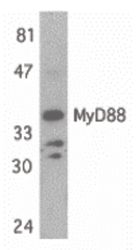
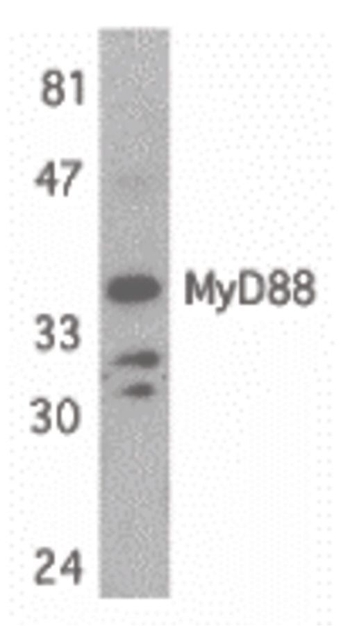
- Western Blot analysis of MyD88 using MyD88 Polyclonal Antibody (Product # PA5-19918) (2 µg/mL) or MyD88 Polyclonal Antibody (Product # PA5-19919) (2 µg/mL). Lysates/proteins at 15 µg per lane. 1 h incubation at RT in 5% NFDM/TBST. Secondary: Goat anti-rabbit IgG HRP conjugate at 1:10,000 dilution.
- Submitted by
- Invitrogen Antibodies (provider)
- Main image

- Experimental details
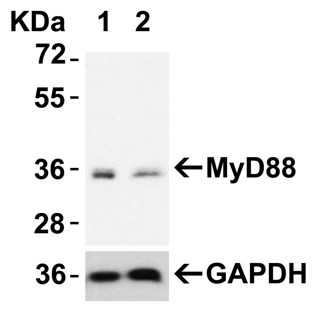
- Western Bloat analysis with MyD88 siRNA Knockdown in HeLa Cells. HeLa cells were transfected with control siRNAs (lane 1) or MyD88 siRNAs (lane 2) Loading: 10 µg of HeLa whole cell lysates per lane. Antibodies: MyD88 Polyclonal Antibody (Product # PA5-19918) (2 µg/mL), 1 h incubation at RT in 5% NFDM/TBST. Secondary: Goat anti-rabbit IgG HRP conjugate at 1:10,000 dilution.
- Submitted by
- Invitrogen Antibodies (provider)
- Main image

- Experimental details
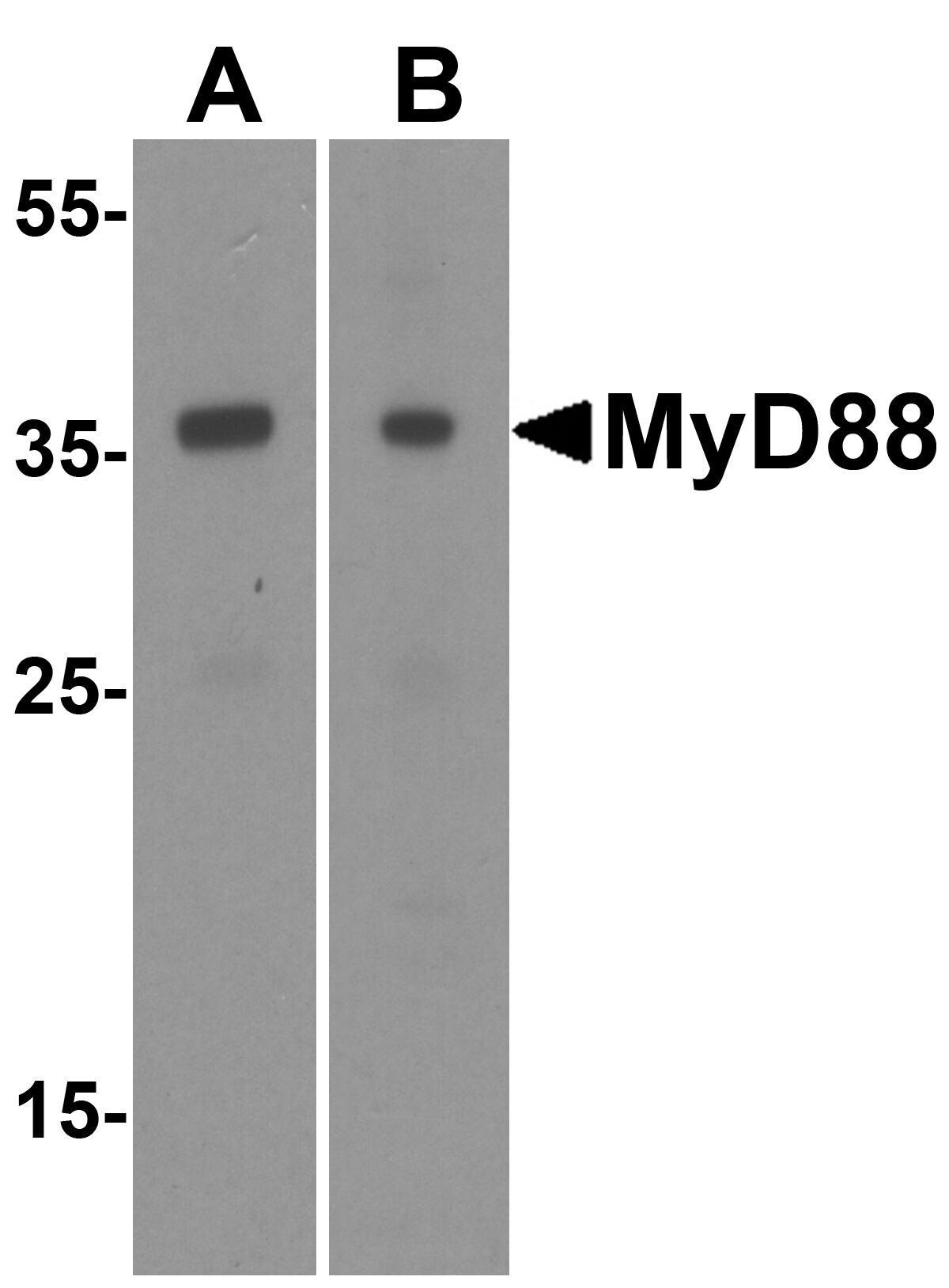
- Western Blot Validation of MyD88 in HeLa (A) and Jurket (B) Cells. Loading: 15 µg of lysates per lane. Antibodies: MyD88 Polyclonal Antibody (Product # PA5-19918) (1 µg/mL) 1 h incubation at RT in 0.05 NFDM/TBST. Secondary: Goat anti-rabbit IgG HRP conjugate at 1:10,000 dilution.
- Submitted by
- Invitrogen Antibodies (provider)
- Main image
- Experimental details
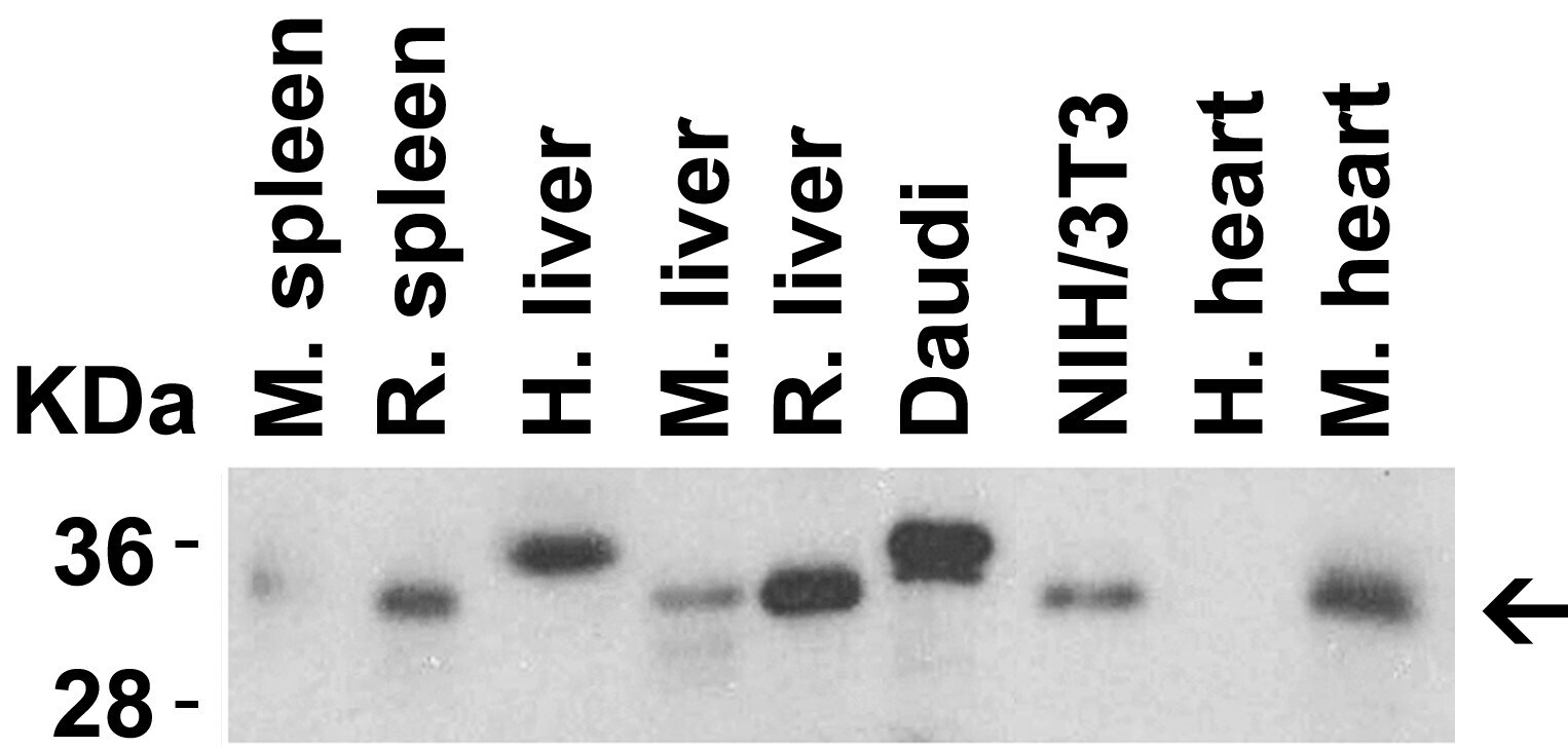
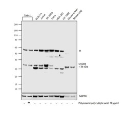
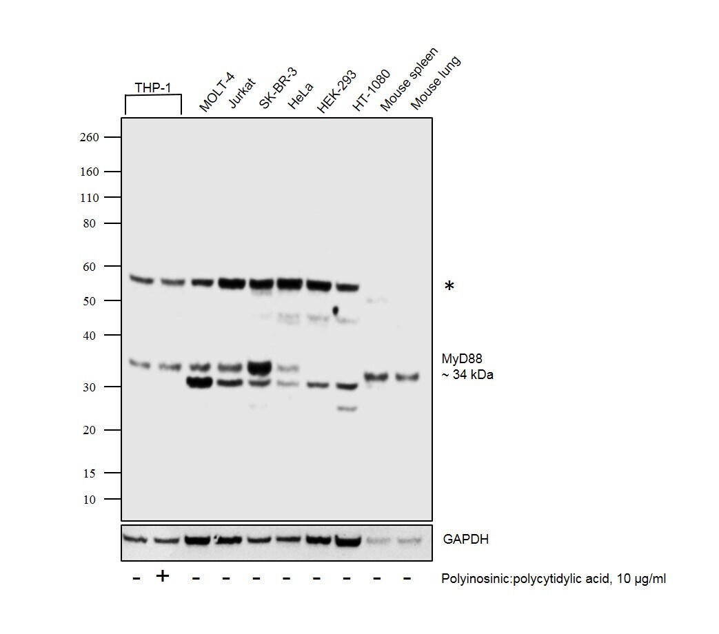
- Western blot was performed using Anti-MyD88 Polyclonal Antibody (Product # PA5-19918) and ~34kDa band corresponding to MyD88 was observed in THP-1, MOLT-4, Jurkat, SK-BR-3, HeLa, Mouse spleen and Mouse lung and not seen in HEK-293 and HT-1080 which are reported to be low expressing cells. Also a lower reported isoform of ~31kDa and an un-characterized band (*) at ~58kDa were also observed in the cell lines and tissues. Whole cell extracts (40 µg lysate) of THP-1 (Lane 1), THP-1 transfected with 10ug/ml of Polyinosinic:Polycytidylic acid (Lane 2), MOLT-4 (Lane 3), Jurkat (Lane 4), SK-BR-3 (Lane 5), HeLa (Lane 6), HEK-293 (Lane 7), HT-1080 (Lane 8), Mouse spleen (Lane 9) and Mouse lung (Lane 10) were electrophoresed using NuPAGE® 4-12% Bis-Tris gel (Product # NP0322BOX). Resolved proteins were then transferred onto a nitrocellulose membrane (Product # IB23001) by iBlot® 2 Dry Blotting System (Product # IB21001).The blot was probed with the primary antibody (1ug/ml) and detected by chemiluminescence with Goat anti-Rabbit IgG (H+L), Superclonal™ Recombinant Secondary Antibody, HRP (Product # A27036, 1:4000 dilution) using the iBright FL 1000 (Product # A32752). Chemiluminescent detection was performed using Novex® ECL Chemiluminescent Substrate Reagent Kit (Product # WP20005)..
Supportive validation
- Submitted by
- Invitrogen Antibodies (provider)
- Main image

- Experimental details
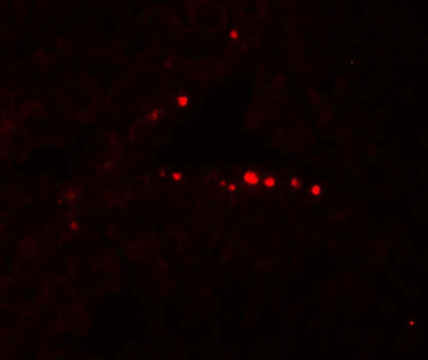
- Immunofluorescent analysis of human testis cells using a MYD88 polyclonal antibody (Product # PA5-19918) at a 20 µg/mL dilution.
- Submitted by
- Invitrogen Antibodies (provider)
- Main image
- Experimental details
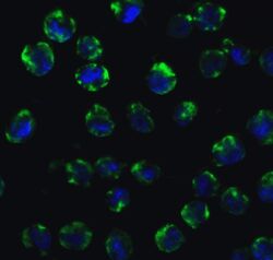
- Immunofluorescent analysis of 4% paraformaldehyde-fixed K562 cells labeling MyD88 with MyD88 Polyclonal Antibody (Product # PA5-19918) at 10 µg/mL, followed by Goat anti-rabbit IgG secondary antibody at 1:500 dilution (green) and DAPI staining (blue).

Supportive validation
- Submitted by
- Invitrogen Antibodies (provider)
- Main image

- Experimental details
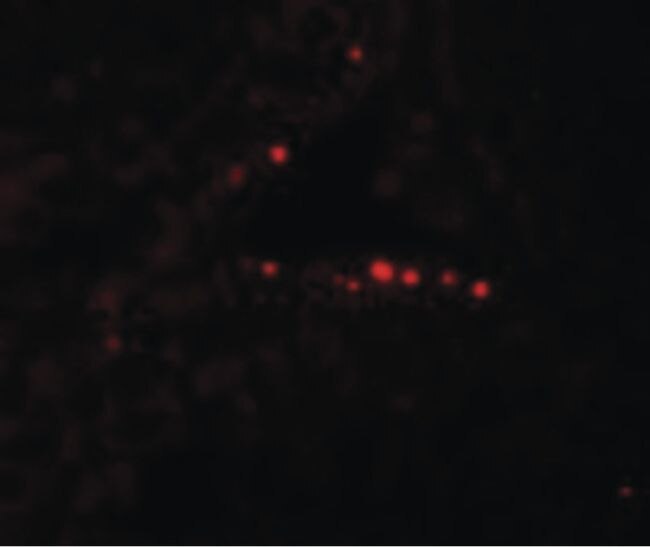
- Immunofluorescent analysis of 4% paraformaldehyde-fixed human testis tissue labeling MyD88 with MyD88 Polyclonal Antibody (Product # PA5-19918) at 20 µg/mL, followed by goat anti-rabbit IgG secondary antibody at 1:500 dilution (red). Image showing nucleus staining on human testis cells.
- Submitted by
- Invitrogen Antibodies (provider)
- Main image

- Experimental details
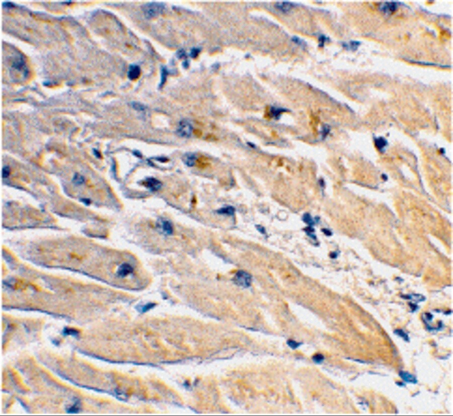
- Immunohistochemical analysis of paraffin-embedded human heart tissue using MyD88 Polyclonal Antibody (Product # PA5-19918) at 2 µg/mL. Tissue was fixed with formaldehyde and blocked with 0.1 serum for 1 h at RT; antigen retrieval was by heat mediation with a citrate buffer (pH6). Samples were incubated with primary antibody overnight at 4˚C. A goat anti-rabbit IgG H&L (HRP) at 1/250 was used as secondary. Counter stained with Hematoxylin.
Supportive validation
- Submitted by
- Invitrogen Antibodies (provider)
- Main image
- Experimental details
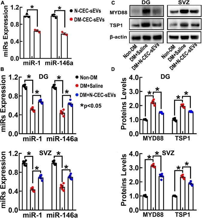
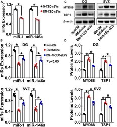
- FIGURE 8 N-CEC-sEV treatment elevates miR-1 and -146a, and reduces MYD88 and TSP1 expression in DG and SVZ tissues isolated from aged-DM rats. Quantitative RT-PCR data show miR-1 and -146a levels in N-CEC-sEVs and DM-CEC-sEVs (A) and in DG and SVZ tissues (B) isolated from age-matched non-DM rats, and aged-DM rats treated with saline and N-CEC-sEVs. Representative Western blots (C) and quantitative data (D) of MYD88 and TSP1 levels in the DG and the SVZ. Data are presented as fold change from the non-DM group. * p < 0.05 versus indicated groups.