Explore
 Validate
 Learn
Learn Western blot
 Immunocytochemistry
Antibody data
- Antibody Data
- Antigen structure
- References [26]
- Comments [0]
- Validations
- Immunocytochemistry [6]
- Immunohistochemistry [3]
- Other assay [4]
Submit
Validation data
Reference
Comment
Report error
- Product number
- MA1-620 - Provider product page

- Provider
- Invitrogen Antibodies
- Product name
- NR3C2 Monoclonal Antibody (H10E4C9F)
- Antibody type
- Monoclonal
- Antigen
- Other
- Description
- MA1-620 detects mineralocorticoid receptor (MR) in human, rat, rabbit and chicken tissues. MA1-620 has been successfully used in Western blot, immunocytochemistry and immunohistochemistry procedures. By Western blot, this antibody detects a 116 kDa protein representing MR in rat heart homogenate. Immunohistochemical staining of MR in rabbit atrium with MA1-620 results in strong staining of myocytes and endothelial cells. MA1-620 cannot be used for immunoprecipitation. In immunohistochemical studies, staining with MA1-620 is blocked by pre-incubating the sample with aldosterone. MA1-620 can also be used in formalin-fixed, paraffin-embedded sections. Due to the extremely low levels of MR expressed in native tissues, it is recommended that enhanced detection systems be used for Western blotting such as enhanced chemiluminescence. MA1-620 blocks the binding of aldosterone since it is directed against the steroid binding domain of the mineralocorticoid receptor. The antibody can also react with the steroid binding domain for aldosterone on other receptors. The MA1-620 immunogen is aldosterone-3. This antibody was produced using the anti-idiotypic method.
- Reactivity
- Human, Rat, Chicken/Avian, Rabbit
- Host
- Mouse
- Isotype
- IgG
- Antibody clone number
- H10E4C9F
- Vial size
- 100 μL
- Concentration
- 1.4 mg/mL
- Storage
- -20°C, Avoid Freeze/Thaw Cycles
Submitted references Loss of central mineralocorticoid or glucocorticoid receptors impacts auditory nerve processing in the cochlea.
Long-Term Programming of CD8 T Cell Immunity by Perinatal Exposure to Glucocorticoids.
Mineralocorticoid receptor is involved in the aldosterone pathway in human red blood cells.
Mineralocorticoid receptor and heat shock protein expression levels in peripheral lymphocytes from war trauma-exposed men with and without PTSD.
Hypothalamic-pituitary-adrenocortical axis hypersensitivity and glucocorticoid receptor expression and function in women with polycystic ovary syndrome.
Mineralocorticoid receptor expression in human venous smooth muscle cells: a potential role for aldosterone signaling in vein graft arterialization.
Aldosterone and mineralocorticoid receptor antagonists modulate elastin and collagen deposition in human skin.
Localization of mineralocorticoid receptors at mammalian synapses.
Cranial irradiation modulates hypothalamic-pituitary-adrenal axis activity and corticosteroid receptor expression in the hippocampus of juvenile rat.
Synergistic induction of osteopontin by aldosterone and inflammatory cytokines in mesangial cells.
Stimulation of testosterone production in rat Leydig cells by aldosterone is mineralocorticoid receptor mediated.
Mineralocorticoid receptor expression in late-gestation ovine fetal lung.
Pituitary expression of type I and type II glucocorticoid receptors during chicken embryonic development and their involvement in growth hormone cell differentiation.
Identification of glucocorticoid receptor domains involved in transrepression of transforming growth factor-beta action.
The mineralocorticoid receptor may compensate for the loss of the glucocorticoid receptor at specific stages of mammary gland development.
The p23 molecular chaperones act at a late step in intracellular receptor action to differentially affect ligand efficacies.
Prerequisite for cardiac aldosterone action. Mineralocorticoid receptor and 11 beta-hydroxysteroid dehydrogenase in the human heart.
Prerequisite for cardiac aldosterone action. Mineralocorticoid receptor and 11 beta-hydroxysteroid dehydrogenase in the human heart.
Human skin as target for aldosterone: coexpression of mineralocorticoid receptors and 11 beta-hydroxysteroid dehydrogenase.
Differential intracellular localization of human mineralocorticosteroid receptor on binding of agonists and antagonists.
Immunohistochemical and biochemical evidence for a cardiovascular mineralocorticoid receptor.
Aldosterone antagonists destabilize the mineralocorticosteroid receptor.
Aldosterone antagonists destabilize the mineralocorticosteroid receptor.
Immunohistochemical localization of renal mineralocorticoid receptor by using an anti-idiotypic antibody that is an internal image of aldosterone.
Immunohistochemical localization of renal mineralocorticoid receptor by using an anti-idiotypic antibody that is an internal image of aldosterone.
Internal image properties of a monoclonal auto-anti-idiotypic antibody and its binding to aldosterone receptors.
Marchetta P, Eckert P, Lukowski R, Ruth P, Singer W, Rüttiger L, Knipper M
iScience 2022 Mar 18;25(3):103981
iScience 2022 Mar 18;25(3):103981
Long-Term Programming of CD8 T Cell Immunity by Perinatal Exposure to Glucocorticoids.
Hong JY, Lim J, Carvalho F, Cho JY, Vaidyanathan B, Yu S, Annicelli C, Ip WKE, Medzhitov R
Cell 2020 Mar 5;180(5):847-861.e15
Cell 2020 Mar 5;180(5):847-861.e15
Mineralocorticoid receptor is involved in the aldosterone pathway in human red blood cells.
Bordin L, Saccardi C, Donà G, Sabbadin C, Andrisani A, Ambrosini G, Plebani M, Brunati AM, Ragazzi E, Gizzo S, Armanini D
American journal of translational research 2016;8(2):314-28
American journal of translational research 2016;8(2):314-28
Mineralocorticoid receptor and heat shock protein expression levels in peripheral lymphocytes from war trauma-exposed men with and without PTSD.
Matić G, Vojnović Milutinović D, Nestorov J, Elaković I, Manitašević Jovanović S, Elzaedi YM, Perišić T, Dunđerski J, Damjanović S, Knežević G, Špirić Ž, Vermetten E, Savić D
Psychiatry research 2014 Feb 28;215(2):379-85
Psychiatry research 2014 Feb 28;215(2):379-85
Hypothalamic-pituitary-adrenocortical axis hypersensitivity and glucocorticoid receptor expression and function in women with polycystic ovary syndrome.
Milutinović DV, Macut D, Božić I, Nestorov J, Damjanović S, Matić G
Experimental and clinical endocrinology & diabetes : official journal, German Society of Endocrinology [and] German Diabetes Association 2011 Nov;119(10):636-43
Experimental and clinical endocrinology & diabetes : official journal, German Society of Endocrinology [and] German Diabetes Association 2011 Nov;119(10):636-43
Mineralocorticoid receptor expression in human venous smooth muscle cells: a potential role for aldosterone signaling in vein graft arterialization.
Bafford R, Sui XX, Park M, Miyahara T, Newfell BG, Jaffe IZ, Romero JR, Adler GK, Williams GH, Khalil RA, Conte MS
American journal of physiology. Heart and circulatory physiology 2011 Jul;301(1):H41-7
American journal of physiology. Heart and circulatory physiology 2011 Jul;301(1):H41-7
Aldosterone and mineralocorticoid receptor antagonists modulate elastin and collagen deposition in human skin.
Mitts TF, Bunda S, Wang Y, Hinek A
The Journal of investigative dermatology 2010 Oct;130(10):2396-406
The Journal of investigative dermatology 2010 Oct;130(10):2396-406
Localization of mineralocorticoid receptors at mammalian synapses.
Prager EM, Brielmaier J, Bergstrom HC, McGuire J, Johnson LR
PloS one 2010 Dec 15;5(12):e14344
PloS one 2010 Dec 15;5(12):e14344
Cranial irradiation modulates hypothalamic-pituitary-adrenal axis activity and corticosteroid receptor expression in the hippocampus of juvenile rat.
Velickovic N, Djordjevic A, Drakulic D, Stanojevic I, Secerov B, Horvat A
General physiology and biophysics 2009;28 Spec No:219-27
General physiology and biophysics 2009;28 Spec No:219-27
Synergistic induction of osteopontin by aldosterone and inflammatory cytokines in mesangial cells.
Gauer S, Hauser IA, Obermüller N, Holzmann Y, Geiger H, Goppelt-Struebe M
Journal of cellular biochemistry 2008 Feb 1;103(2):615-23
Journal of cellular biochemistry 2008 Feb 1;103(2):615-23
Stimulation of testosterone production in rat Leydig cells by aldosterone is mineralocorticoid receptor mediated.
Ge RS, Dong Q, Sottas CM, Latif SA, Morris DJ, Hardy MP
Molecular and cellular endocrinology 2005 Nov 24;243(1-2):35-42
Molecular and cellular endocrinology 2005 Nov 24;243(1-2):35-42
Mineralocorticoid receptor expression in late-gestation ovine fetal lung.
Keller-Wood M, Wood CE, Hua Y, Zhang D
Journal of the Society for Gynecologic Investigation 2005 Feb;12(2):84-91
Journal of the Society for Gynecologic Investigation 2005 Feb;12(2):84-91
Pituitary expression of type I and type II glucocorticoid receptors during chicken embryonic development and their involvement in growth hormone cell differentiation.
Bossis I, Nishimura S, Muchow M, Porter TE
Endocrinology 2004 Jul;145(7):3523-31
Endocrinology 2004 Jul;145(7):3523-31
Identification of glucocorticoid receptor domains involved in transrepression of transforming growth factor-beta action.
Li G, Wang S, Gelehrter TD
The Journal of biological chemistry 2003 Oct 24;278(43):41779-88
The Journal of biological chemistry 2003 Oct 24;278(43):41779-88
The mineralocorticoid receptor may compensate for the loss of the glucocorticoid receptor at specific stages of mammary gland development.
Kingsley-Kallesen M, Mukhopadhyay SS, Wyszomierski SL, Schanler S, Schütz G, Rosen JM
Molecular endocrinology (Baltimore, Md.) 2002 Sep;16(9):2008-18
Molecular endocrinology (Baltimore, Md.) 2002 Sep;16(9):2008-18
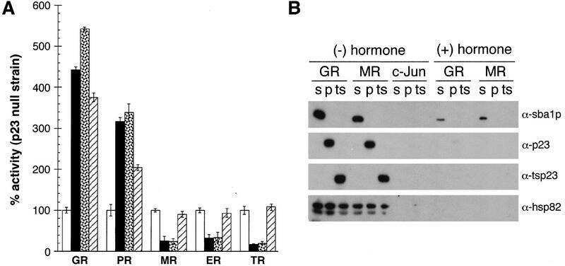
The p23 molecular chaperones act at a late step in intracellular receptor action to differentially affect ligand efficacies.
Freeman BC, Felts SJ, Toft DO, Yamamoto KR
Genes & development 2000 Feb 15;14(4):422-34
Genes & development 2000 Feb 15;14(4):422-34
Prerequisite for cardiac aldosterone action. Mineralocorticoid receptor and 11 beta-hydroxysteroid dehydrogenase in the human heart.
Lombès M, Alfaidy N, Eugene E, Lessana A, Farman N, Bonvalet JP
Circulation 1995 Jul 15;92(2):175-82
Circulation 1995 Jul 15;92(2):175-82
Prerequisite for cardiac aldosterone action. Mineralocorticoid receptor and 11 beta-hydroxysteroid dehydrogenase in the human heart.
Lombès M, Alfaidy N, Eugene E, Lessana A, Farman N, Bonvalet JP
Circulation 1995 Jul 15;92(2):175-82
Circulation 1995 Jul 15;92(2):175-82
Human skin as target for aldosterone: coexpression of mineralocorticoid receptors and 11 beta-hydroxysteroid dehydrogenase.
Kenouch S, Lombes M, Delahaye F, Eugene E, Bonvalet JP, Farman N
The Journal of clinical endocrinology and metabolism 1994 Nov;79(5):1334-41
The Journal of clinical endocrinology and metabolism 1994 Nov;79(5):1334-41
Differential intracellular localization of human mineralocorticosteroid receptor on binding of agonists and antagonists.
Lombès M, Binart N, Delahaye F, Baulieu EE, Rafestin-Oblin ME
The Biochemical journal 1994 Aug 15;302 ( Pt 1):191-7
The Biochemical journal 1994 Aug 15;302 ( Pt 1):191-7
Immunohistochemical and biochemical evidence for a cardiovascular mineralocorticoid receptor.
Lombès M, Oblin ME, Gasc JM, Baulieu EE, Farman N, Bonvalet JP
Circulation research 1992 Sep;71(3):503-10
Circulation research 1992 Sep;71(3):503-10
Aldosterone antagonists destabilize the mineralocorticosteroid receptor.
Couette B, Lombes M, Baulieu EE, Rafestin-Oblin ME
The Biochemical journal 1992 Mar 15;282 ( Pt 3)(Pt 3):697-702
The Biochemical journal 1992 Mar 15;282 ( Pt 3)(Pt 3):697-702
Aldosterone antagonists destabilize the mineralocorticosteroid receptor.
Couette B, Lombes M, Baulieu EE, Rafestin-Oblin ME
The Biochemical journal 1992 Mar 15;282 ( Pt 3):697-702
The Biochemical journal 1992 Mar 15;282 ( Pt 3):697-702
Immunohistochemical localization of renal mineralocorticoid receptor by using an anti-idiotypic antibody that is an internal image of aldosterone.
Lombès M, Farman N, Oblin ME, Baulieu EE, Bonvalet JP, Erlanger BF, Gasc JM
Proceedings of the National Academy of Sciences of the United States of America 1990 Feb;87(3):1086-8
Proceedings of the National Academy of Sciences of the United States of America 1990 Feb;87(3):1086-8
Internal image properties of a monoclonal auto-anti-idiotypic antibody and its binding to aldosterone receptors.
Lombes M, Edelman IS, Erlanger BF
The Journal of biological chemistry 1989 Feb 15;264(5):2528-36
The Journal of biological chemistry 1989 Feb 15;264(5):2528-36
No comments: Submit comment
Supportive validation
- Submitted by
- Invitrogen Antibodies (provider)
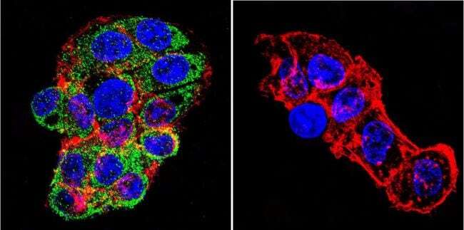
- Main image

- Experimental details
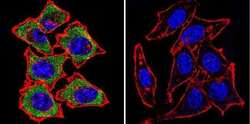
- Immunofluorescent analysis of Mineralocorticoid Receptor using Mineralocorticoid Receptor Monoclonal antibody (H10E4C9F) (Product # MA1-620) shows staining in HEK293 cells. Mineralocorticoid Receptor staining (green), F-Actin staining with Phalloidin (red) and nuclei with DAPI (blue) is shown. Cells were grown on chamber slides and fixed with formaldehyde prior to staining. Cells were probed without (control) or with or an antibody recognizing Mineralocorticoid Receptor (Product # MA1-620) at a dilution of 1:20-1:200 over night at 4 °C, washed with PBS and incubated with a DyLight-488 conjugated secondary antibody (Product # 35552 for GAR, Product # 35503 for GAM). Images were taken at 60X magnification.

- Submitted by
- Invitrogen Antibodies (provider)
- Main image
- Experimental details
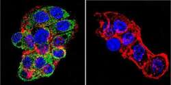
- Immunofluorescent analysis of Mineralocorticoid Receptor using Mineralocorticoid Receptor Monoclonal antibody (H10E4C9F) (Product # MA1-620) shows staining in HeLa cells. Mineralocorticoid Receptor staining (green), F-Actin staining with Phalloidin (red) and nuclei with DAPI (blue) is shown. Cells were grown on chamber slides and fixed with formaldehyde prior to staining. Cells were probed without (control) or with or an antibody recognizing Mineralocorticoid Receptor (Product # MA1-620) at a dilution of 1:20-1:200 over night at 4 °C, washed with PBS and incubated with a DyLight-488 conjugated secondary antibody (Product # 35552 for GAR, Product # 35503 for GAM). Images were taken at 60X magnification.

- Submitted by
- Invitrogen Antibodies (provider)
- Main image
- Experimental details
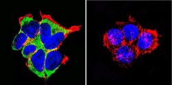
- Immunofluorescent analysis of Mineralocorticoid Receptor using Mineralocorticoid Receptor Monoclonal antibody (H10E4C9F) (Product # MA1-620) shows staining in HepG2 cells. Mineralocorticoid Receptor staining (green), F-Actin staining with Phalloidin (red) and nuclei with DAPI (blue) is shown. Cells were grown on chamber slides and fixed with formaldehyde prior to staining. Cells were probed without (control) or with or an antibody recognizing Mineralocorticoid Receptor (Product # MA1-620) at a dilution of 1:20-1:200 over night at 4 °C, washed with PBS and incubated with a DyLight-488 conjugated secondary antibody (Product # 35552 for GAR, Product # 35503 for GAM). Images were taken at 60X magnification.

- Submitted by
- Invitrogen Antibodies (provider)
- Main image
- Experimental details
- Immunofluorescent analysis of Mineralocorticoid Receptor using Mineralocorticoid Receptor Monoclonal antibody (H10E4C9F) (Product # MA1-620) shows staining in HEK293 cells. Mineralocorticoid Receptor staining (green), F-Actin staining with Phalloidin (red) and nuclei with DAPI (blue) is shown. Cells were grown on chamber slides and fixed with formaldehyde prior to staining. Cells were probed without (control) or with or an antibody recognizing Mineralocorticoid Receptor (Product # MA1-620) at a dilution of 1:20-1:200 over night at 4 °C, washed with PBS and incubated with a DyLight-488 conjugated secondary antibody (Product # 35552 for GAR, Product # 35503 for GAM). Images were taken at 60X magnification.

- Submitted by
- Invitrogen Antibodies (provider)
- Main image

- Experimental details
- Immunofluorescent analysis of Mineralocorticoid Receptor using Mineralocorticoid Receptor Monoclonal antibody (H10E4C9F) (Product # MA1-620) shows staining in HeLa cells. Mineralocorticoid Receptor staining (green), F-Actin staining with Phalloidin (red) and nuclei with DAPI (blue) is shown. Cells were grown on chamber slides and fixed with formaldehyde prior to staining. Cells were probed without (control) or with or an antibody recognizing Mineralocorticoid Receptor (Product # MA1-620) at a dilution of 1:20-1:200 over night at 4 °C, washed with PBS and incubated with a DyLight-488 conjugated secondary antibody (Product # 35552 for GAR, Product # 35503 for GAM). Images were taken at 60X magnification.

- Submitted by
- Invitrogen Antibodies (provider)
- Main image

- Experimental details
- Immunofluorescent analysis of Mineralocorticoid Receptor using Mineralocorticoid Receptor Monoclonal antibody (H10E4C9F) (Product # MA1-620) shows staining in HepG2 cells. Mineralocorticoid Receptor staining (green), F-Actin staining with Phalloidin (red) and nuclei with DAPI (blue) is shown. Cells were grown on chamber slides and fixed with formaldehyde prior to staining. Cells were probed without (control) or with or an antibody recognizing Mineralocorticoid Receptor (Product # MA1-620) at a dilution of 1:20-1:200 over night at 4 °C, washed with PBS and incubated with a DyLight-488 conjugated secondary antibody (Product # 35552 for GAR, Product # 35503 for GAM). Images were taken at 60X magnification.
Supportive validation
- Submitted by
- Invitrogen Antibodies (provider)
- Main image
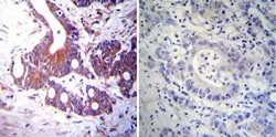
- Experimental details
- Immunohistochemistry was performed on cancer biopsies of deparaffinized Human colon carcinoma tissues. To expose target proteins, heat induced antigen retrieval was performed using 10mM sodium citrate (pH6.0) buffer, microwaved for 8-15 minutes. Following antigen retrieval tissues were blocked in 3% BSA-PBS for 30 minutes at room temperature. Tissues were then probed at a dilution of 1:100 with a mouse monoclonal antibody recognizing Mineralocorticoid Receptor (Product # MA1-620) or without primary antibody (negative control) overnight at 4°C in a humidified chamber. Tissues were washed extensively with PBST and endogenous peroxidase activity was quenched with a peroxidase suppressor. Detection was performed using a biotin-conjugated secondary antibody and SA-HRP, followed by colorimetric detection using DAB. Tissues were counterstained with hematoxylin and prepped for mounting.
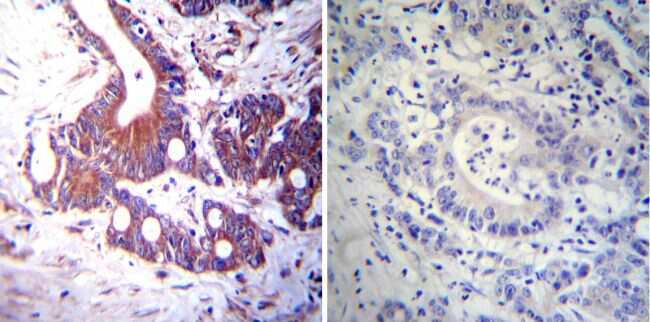
- Submitted by
- Invitrogen Antibodies (provider)
- Main image
- Experimental details
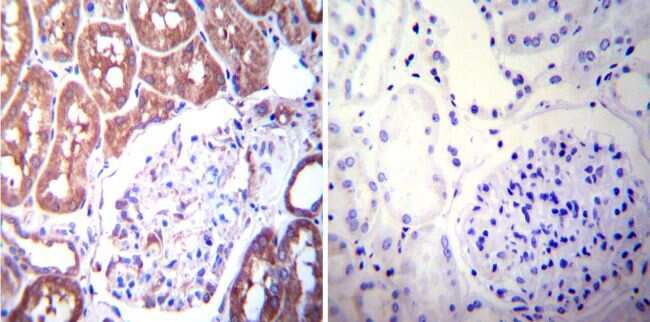
- Immunohistochemistry was performed on normal deparaffinized Human kidney tissue tissues. To expose target proteins, heat induced antigen retrieval was performed using 10mM sodium citrate (pH6.0) buffer, microwaved for 8-15 minutes. Following antigen retrieval tissues were blocked in 3% BSA-PBS for 30 minutes at room temperature. Tissues were then probed at a dilution of 1:200 with a mouse monoclonal antibody recognizing Mineralocorticoid Receptor (Product # MA1-620) or without primary antibody (negative control) overnight at 4°C in a humidified chamber. Tissues were washed extensively with PBST and endogenous peroxidase activity was quenched with a peroxidase suppressor. Detection was performed using a biotin-conjugated secondary antibody and SA-HRP, followed by colorimetric detection using DAB. Tissues were counterstained with hematoxylin and prepped for mounting.
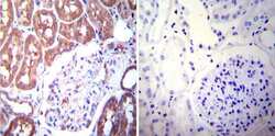
- Submitted by
- Invitrogen Antibodies (provider)
- Main image
- Experimental details
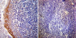
- Immunohistochemistry was performed on normal deparaffinized Human tonsil tissue tissues. To expose target proteins, heat induced antigen retrieval was performed using 10mM sodium citrate (pH6.0) buffer, microwaved for 8-15 minutes. Following antigen retrieval tissues were blocked in 3% BSA-PBS for 30 minutes at room temperature. Tissues were then probed at a dilution of 1:100 with a mouse monoclonal antibody recognizing Mineralocorticoid Receptor (Product # MA1-620) or without primary antibody (negative control) overnight at 4°C in a humidified chamber. Tissues were washed extensively with PBST and endogenous peroxidase activity was quenched with a peroxidase suppressor. Detection was performed using a biotin-conjugated secondary antibody and SA-HRP, followed by colorimetric detection using DAB. Tissues were counterstained with hematoxylin and prepped for mounting.

Supportive validation
- Submitted by
- Invitrogen Antibodies (provider)
- Main image
- Experimental details
- NULL

- Submitted by
- Invitrogen Antibodies (provider)
- Main image
- Experimental details
- NULL
- Submitted by
- Invitrogen Antibodies (provider)
- Main image
- Experimental details
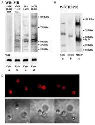
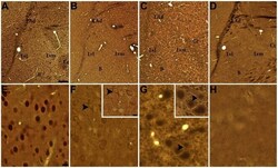
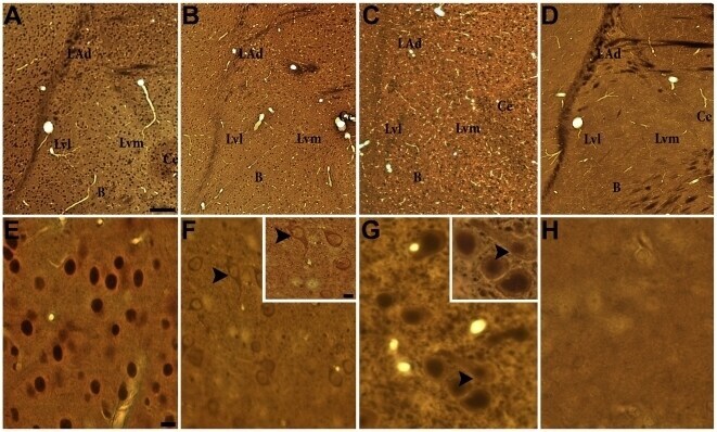
- Figure 2 GR and MR labeling in the rat amygdala. (A & E) GR-ir neurons are distributed throughout the amygdala predominately labeling the nucleus. (B & F) MR-ir (MA1-620) neurons are distributed throughout the amygdala but immunoreactivity is not as dense as GR-ir. Large proximal dendrites showed immunoreactivity (F inset). (C & G) MR-ir (rMR1-18 1D5) neurons are distributed in amygdala nuclei and cell membrane but do not appear as densely labeled in perikaryon (G inset). (D & H) Control section shows little immunoreactivity of cells in amygdala or surrounding nuclei. Scale bars = 200 um for A-D, 10 um for D-G and 5 um for F & G insets. Amygdala nuclei subdivisions B = basal, Ce = central, Lad = lateral (dorsal division), Lvl = lateral (ventral-lateral), Lvm = lateral (ventral-medial).
- Submitted by
- Invitrogen Antibodies (provider)
- Main image

- Experimental details
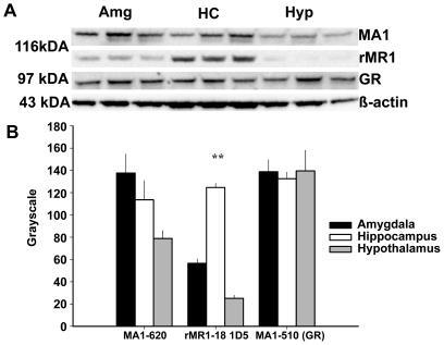
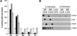
- Figure 1 Immunoblots of MR and GR in amygdala (Amg), hippocampal (HC) and hypothalamic (Hyp) nuclei. As indicated by the representative immunoblots standardized to beta-Actin (A) there was a significant difference in relative density of MR between brain nuclei (B). GR was equally distributed in all brain regions. (** denotes p's