Explore
 Validate
 Learn
Learn Western blot
 Immunocytochemistry
Antibody data
- Antibody Data
- Antigen structure
- References [1]
- Comments [0]
- Validations
- Western blot [1]
Submit
Validation data
Reference
Comment
Report error
- Product number
- M01019-2 - Provider product page

- Provider
- Boster Biological Technology
- Product name
- Anti-GPX1 Antibody Picoband™ (monoclonal, 8B10)
- Antibody type
- Monoclonal
- Description
- Mouse IgG monoclonal antibody for GPX1 detection. Tested with WB, IHC-P, ICC/IF, FCM in Human;Mouse;Rat.
- Reactivity
- Human, Mouse, Rat
- Host
- Mouse
- Isotype
- IgG
- Antibody clone number
- 8B10
- Vial size
- 100μg/vial
- Concentration
- 0.5-1mg/ml, actual concentration vary by lot. Use suggested dilution ratio to decide dilution procedure.
- Storage
- At -20°C for one year. After reconstitution, at 4°C for one month. It can also be aliquoted and stored frozen at -20°C for a longer time. Avoid repeated freezing and thawing.
- Handling
- Add 0.2ml of distilled water will yield a concentration of 500μg/ml.
Submitted references Injectable hydrogel with selenium nanoparticles delivery for sustained glutathione peroxidase activation and enhanced osteoarthritis therapeutics.
Hu W, Yao X, Li Y, Li J, Zhang J, Zou Z, Kang F, Dong S
Materials today. Bio 2023 Dec;23:100864
Materials today. Bio 2023 Dec;23:100864
No comments: Submit comment
Supportive validation
- Submitted by
- Boster Biological Technology (provider)
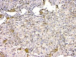
- Main image
- Experimental details
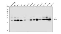
- Western blot analysis of GPX1 using anti-GPX1 antibody (M01019-2). Electrophoresis was performed on a 5-20% SDS-PAGE gel at 70V (Stacking gel) / 90V (Resolving gel) for 2-3 hours. The sample well of each lane was loaded with 50ug of sample under reducing conditions. Lane 1: human placenta tissue lysates, Lane 2: human U-87 whole cell lysates, Lane 3: human THP-1 whole cell lysates, Lane 4: human U-937 whole cell lysates, Lane 5: human HepG2 whole cell lysates, Lane 6: rat brain tissue lysates, Lane 7: rat thymus tissue lysates, Lane 8: rat lung tissue lysates, Lane 9: rat liver tissue lysates, Lane 10: mouse brain tissue lysates, Lane 11: mouse thymus tissue lysates, Lane 12: mouse lung tissue lysates, Lane 13: mouse liver tissue lysates. After Electrophoresis, proteins were transferred to a Nitrocellulose membrane at 150mA for 50-90 minutes. Blocked the membrane with 5% Non-fat Milk/ TBS for 1.5 hour at RT. The membrane was incubated with mouse anti-GPX1 antigen affinity purified monoclonal antibody (Catalog # M01019-2) at 0.5 μg/mL overnight at 4°C, then washed with TBS-0.1%Tween 3 times with 5 minutes each and probed with a goat anti-mouse IgG-HRP secondary antibody at a dilution of 1:10000 for 1.5 hour at RT. The signal is developed using an Enhanced Chemiluminescent detection (ECL) kit (Catalog # EK1001) with Tanon 5200 system. A specific band was detected for GPX1 at approximately 22KD. The expected band size for GPX1 is at 22KD.
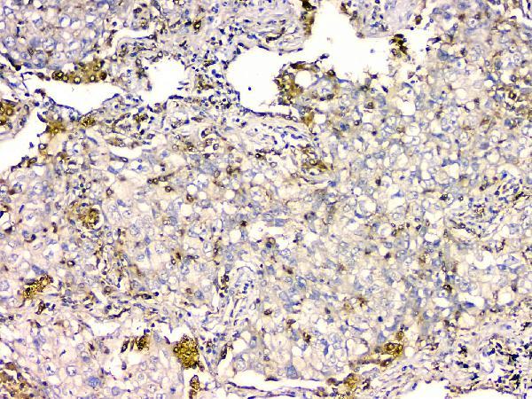
- Additional image