Explore
 Validate
 Learn
Learn Western blot
 Immunohistochemistry
Antibody data
- Antibody Data
- Antigen structure
- References [3]
- Comments [0]
- Validations
- Western blot [4]
- Immunocytochemistry [1]
Submit
Validation data
Reference
Comment
Report error
- Product number
- PA5-30593 - Provider product page

- Provider
- Invitrogen Antibodies
- Product name
- Anti-GPX1 Polyclonal Antibody
- Antibody type
- Polyclonal
- Antigen
- Synthetic peptide
- Description
- Recommended positive controls: Molt-4, Raji, mouse liver, GL261, C8D30, NIH-3T3, Raw264.7, C2C12. Predicted reactivity: Rhesus Monkey (100%), Chimpanzee (100%). Store product as a concentrated solution. Centrifuge briefly prior to opening the vial.
- Reactivity
- Human, Mouse, Rat
- Host
- Rabbit
- Isotype
- IgG
- Vial size
- 100 µL
- Concentration
- 3.14 mg/mL
- Storage
- Store at 4°C short term. For long term storage, store at -20°C, avoiding freeze/thaw cycles.
Submitted references PDGF-BB Preserves Mitochondrial Morphology, Attenuates ROS Production, and Upregulates Neuroglobin in an Astrocytic Model Under Rotenone Insult.
Diabetes-induced hepatic oxidative stress: a new pathogenic role for glycated albumin.
Role of tRNA selenocysteine 1 associated protein 1 in the proliferation and apoptosis of cardiomyocyte‑like H9c2 cells.
Cabezas R, Vega-Vela NE, González-Sanmiguel J, González J, Esquinas P, Echeverria V, Barreto GE
Molecular neurobiology 2018 Apr;55(4):3085-3095
Molecular neurobiology 2018 Apr;55(4):3085-3095
Diabetes-induced hepatic oxidative stress: a new pathogenic role for glycated albumin.
Patche J, Girard D, Catan A, Boyer F, Dobi A, Planesse C, Diotel N, Guerin-Dubourg A, Baret P, Bravo SB, Paradela-Dobarro B, Álvarez E, Essop MF, Meilhac O, Bourdon E, Rondeau P
Free radical biology & medicine 2017 Jan;102:133-148
Free radical biology & medicine 2017 Jan;102:133-148
Role of tRNA selenocysteine 1 associated protein 1 in the proliferation and apoptosis of cardiomyocyte‑like H9c2 cells.
Li MD, Cheng WP, Shi MX, Ge TD, Zheng XL, Wu DY, Hu XY, Luo JC, Li FL, Li H
Molecular medicine reports 2017 Feb;15(2):988-994
Molecular medicine reports 2017 Feb;15(2):988-994
No comments: Submit comment
Supportive validation
- Submitted by
- Invitrogen Antibodies (provider)
- Main image
- Experimental details
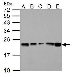
- Western blot analysis of GPX1 using A) 30 µg GL261 whole cell lysate (B) 30 µg C8D30 whole cell lysate (C) 30 µg NIH-3T3 whole cell lysate (D) 30 µg Raw264.7 whole cell lysate and E) 30 µg C2C12 whole cell lysate. Samples were loaded onto a 12% SDS-PAGE gel and probed with a GPX1 polyclonal antibody (Product # PA5-30593) at a dilution of 1:2000.
- Submitted by
- Invitrogen Antibodies (provider)
- Main image
- Experimental details
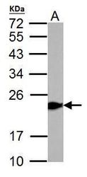
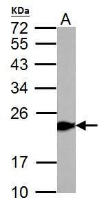
- Western blot analysis of GPX1 using 50 µg rat liver lysate. Samples were loaded onto a 12% SDS-PAGE gel and probed with a GPX1 polyclonal antibody (Product # PA5-30593) at a dilution of 1:10,000.
- Submitted by
- Invitrogen Antibodies (provider)
- Main image

- Experimental details
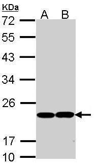
- Western blot analysis of GPX1 using 30 µg of A) MOLT4 and B) Raji lysate. Samples were loaded onto a 12% SDS-PAGE gel and probed with a GPX1 polyclonal antibody (Product # PA5-30593) at a dilution of 1:1000.
- Submitted by
- Invitrogen Antibodies (provider)
- Main image
- Experimental details
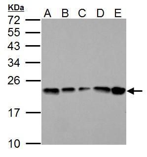
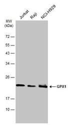
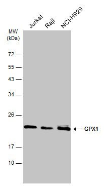
- Western Blot analysis of GPX1 was performed by separating 30 µg of various whole cell extracts by 12% SDS-PAGE. Proteins were transferred to a membrane and probed with a GPX1 Polyclonal Antibody (Product # PA5-30593) at a dilution of 1:1000 and a HRP-conjugated anti-rabbit IgG secondary antibody.
Supportive validation
- Submitted by
- Invitrogen Antibodies (provider)
- Main image
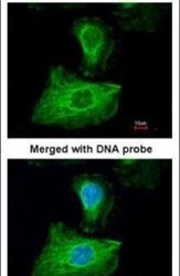
- Experimental details
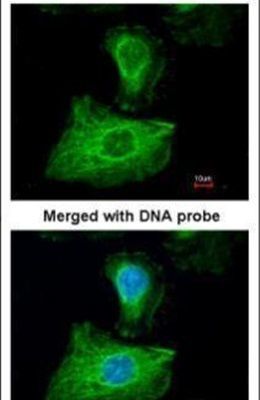
- Immunofluorescent analysis of GPX1 in paraformaldehyde-fixed HeLa cells using a GPX1 polyclonal antibody (Product # PA5-30593) at a 1:200 dilution.