Explore
 Validate
 Learn
LearnHPA019127
antibody from Atlas Antibodies
Targeting: SMARCB1
BAF47, hSNFS, Ini1, PPP1R144, RDT, Sfh1p, SNF5, SNF5L1, Snr1
 Western blot
 Immunohistochemistry
 Chromatin Immunoprecipitation
Antibody data
- Antibody Data
- Antigen structure
- References [0]
- Comments [0]
- Validations
- Western blot [1]
- Chromatin Immunoprecipitation [1]
Submit
Validation data
Reference
Comment
Report error
- Product number
- HPA019127 - Provider product page

- Provider
- Atlas Antibodies
- Proper citation
- Atlas Antibodies Cat#HPA019127, RRID:AB_1851165
- Product name
- Anti-SMARCB1
- Antibody type
- Polyclonal
- Description
- Polyclonal Antibody against Human SMARCB1, Gene description: SWI/SNF related, matrix associated, actin dependent regulator of chromatin, subfamily b, member 1, Alternative Gene Names: BAF47, hSNFS, Ini1, PPP1R144, RDT, Sfh1p, SNF5, SNF5L1, Snr1, Validated applications: WB, IHC, ChIP, Uniprot ID: Q12824, Storage: Store at +4°C for short term storage. Long time storage is recommended at -20°C.
- Reactivity
- Human, Mouse, Rat
- Host
- Rabbit
- Conjugate
- Unconjugated
- Isotype
- IgG
- Vial size
- 100 µl
- Concentration
- 0.1 mg/ml
- Storage
- Store at +4°C for short term storage. Long time storage is recommended at -20°C.
- Handling
- The antibody solution should be gently mixed before use.
No comments: Submit comment
Enhanced validation
- Submitted by
- Atlas Antibodies (provider)
- Enhanced method
- Genetic validation
- Main image

- Experimental details
- Western blot analysis in HEK293 cells transfected with control siRNA, target specific siRNA probe #1 and #2, using Anti-SMARCB1 antibody. Remaining relative intensity is presented. Loading control: Anti-GAPDH.
- Sample type
- Human
- Protocol
- Protocol

Supportive validation
- Submitted by
- Atlas Antibodies (provider)
- Main image
- Experimental details
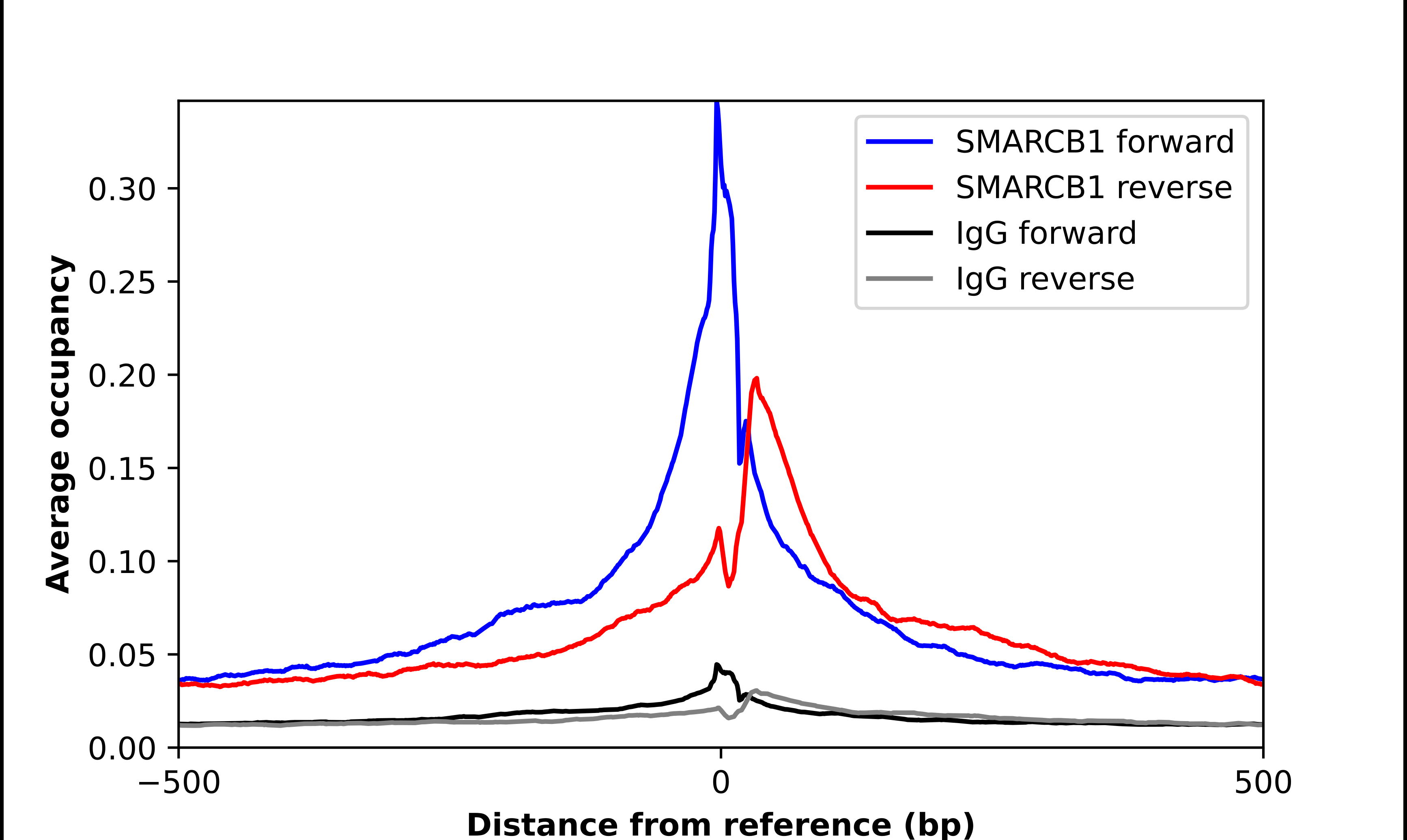
- ChIP-Exo-Seq composite graph for Anti-SMARCB1 (HPA019127, Lot 000000608) tested in K562 cells. Strand-specific reads (blue: forward, red: reverse) and IgG controls (black: forward, grey: reverse) are plotted against the distance from a composite set of reference binding sites. The antibody exhibits robust target enrichment compared to a non-specific IgG control and precisely reveals its structural organization around the binding site. Data generated by Prof. B. F. Pugh´s Lab at Cornell University.