 Explore
 Validate
 Learn
Learn Western blot
Antibody data
- Antibody Data
- Antigen structure
- References [1]
- Comments [0]
- Validations
- Western blot [1]
- Immunocytochemistry [1]
Submit
Validation data
Reference
Comment
Report error
- Product number
- 701985 - Provider product page

- Provider
- Invitrogen Antibodies
- Product name
- TrkC Recombinant Rabbit Monoclonal Antibody (7H3L20)
- Antibody type
- Monoclonal
- Antigen
- Other
- Description
- This antibody is predicted to react with Monkey, Mouse and Rat.
- Antibody clone number
- 7H3L20
- Concentration
- 0.5 mg/mL
Submitted references In vivo survival and differentiation of Friedreich ataxia iPSC-derived sensory neurons transplanted in the adult dorsal root ganglia.
Viventi S, Frausin S, Howden SE, Lim SY, Finol-Urdaneta RK, McArthur JR, Abu-Bonsrah KD, Ng W, Ivanusic J, Thompson L, Dottori M
Stem cells translational medicine 2021 Aug;10(8):1157-1169
Stem cells translational medicine 2021 Aug;10(8):1157-1169
No comments: Submit comment
Supportive validation
- Submitted by
- Invitrogen Antibodies (provider)
- Main image

- Experimental details
- Western blot analysis was performed on whole cell extracts (30 µg lysate) of K562 (Lane 1), K562 treated with PMA (200nM for 20 min) (Lane 2), SH-SY5Y (Lane 3), SH-SY5Y treated with PMA (200nM for 20 min) (Lane 4), A549 (Lane 5), A549 treated with PMA (200nM for 20 min) (Lane 6), NTERA-2 (Lane 7) and NTERA-2 treated with PMA (Lane 8). The blots were probed with TrkC Recombinant Rabbit Monoclonal Antibody (Product # 701985, 1-2 µg/mL) and detected by chemiluminescence using Goat anti-Rabbit IgG (H+L) Superclonal™ Secondary Antibody, HRP conjugate (Product # A27036, 0.4 µg/mL, 1:2500 dilution). A 94 kDa band corresponding to TrkC was observed in a treatment dependent manner in most cell lines tested. Known quantity of protein samples were electrophoresed using Novex® NuPAGE® 4-12% Bis-Tris gel (Product # NP0321BOX), XCell SureLock™ Electrophoresis System (Product # EI0002) and Novex® Sharp Pre-Stained Protein Standard (Product # LC5800). Resolved proteins were then transferred onto a nitrocellulose membrane with iBlot® Dry Blotting System (Product # IB21001). The membrane was probed with the relevant primary and secondary Antibody following blocking with 5% skimmed milk. Chemiluminescent detection was performed using Pierce™ ECL Western blotting Substrate (Product # 32106).

Supportive validation
- Submitted by
- Invitrogen Antibodies (provider)
- Main image
- Experimental details
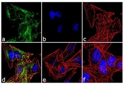
- For immunofluorescence analysis, retinoic acid (10 uM, 72 hours) treated SH-SY5Y cells were fixed and permeabilized for detection of endogenous TrkC using Anti-TrkC Recombinant Rabbit Monoclonal Antibody (Product # 701985, 2 µg/mL) and labeled with Goat anti-Rabbit IgG (H+L) Superclonal™ Secondary Antibody, Alexa Fluor® 488 conjugate (Product # A27034, 1:2000). Panel a) shows representative cells that were stained for detection and localization of TrkC protein (green), Panel b) is stained for nuclei (blue) using SlowFade® Gold Antifade Mountant with DAPI (Product # S36938). Panel c) represents cytoskeletal F-actin staining using Alexa Fluor® 555 Rhodamine Phalloidin (Product # R415, 1:300). Panel d) is a composite image of Panels a, b and c clearly demonstrating membrane localization of TrkC. Panel e) shows untreated cells with no signal. Panel f) represents control cells with no primary antibody to assess background. The images were captured at 60X magnification.
