Explore
 Validate
 Learn
Learn Western blot
Antibody data
- Antibody Data
- Antigen structure
- References [0]
- Comments [0]
- Validations
- Western blot [2]
- Immunocytochemistry [2]
- Immunohistochemistry [1]
Submit
Validation data
Reference
Comment
Report error
- Product number
- ANT-020-25UL - Provider product page

- Provider
- Invitrogen Antibodies
- Product name
- TrkC (extracellular) Polyclonal Antibody
- Antibody type
- Polyclonal
- Antigen
- Other
- Reactivity
- Human, Mouse, Rat
- Host
- Rabbit
- Isotype
- IgG
- Vial size
- 25 µL
- Concentration
- 0.8 mg/mL
- Storage
- -20° C, Avoid Freeze/Thaw Cycles
No comments: Submit comment
Supportive validation
- Submitted by
- Invitrogen Antibodies (provider)
- Main image
- Experimental details
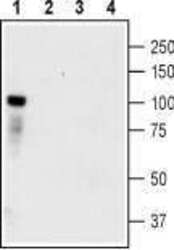
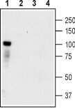
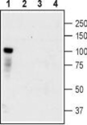
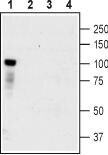
- Western blot analysis of HEK-TrkC transfected cells (lanes 1 and 3) and HEK untransfected cells (lanes 2 and 4) lysates: - 1,2. Anti-TrkC (extracellular) Antibody (#ANT-020), (1:400).3,4. Anti-TrkC (extracellular) Antibody , preincubated with TrkC (extracellular) Blocking Peptide (#BLP-NT020).
- Submitted by
- Invitrogen Antibodies (provider)
- Main image
- Experimental details
- Western blot analysis of HEK-TrkC transfected cells (lanes 1 and 3) and HEK untransfected cells (lanes 2 and 4) lysates: - 1,2. Anti-TrkC (extracellular) Antibody (#ANT-020), (1:400).3,4. Anti-TrkC (extracellular) Antibody , preincubated with TrkC (extracellular) Blocking Peptide (#BLP-NT020).
Supportive validation
- Submitted by
- Invitrogen Antibodies (provider)
- Main image

- Experimental details
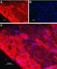
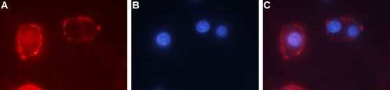
- Expression of TrkC in rat PC12 cells - Cell surface detection of TrkC in live intact ratpheochromocytoma PC12 cells. A. Extracellular staining of cells using Anti-TrkC (extracellular) Antibody (#ANT-020), (1:50) followed by goat Anti-rabbit-AlexaFluor-594 secondary Antibody (red). B. Nuclear staining using the cell permeable dye Hoechst 33342 (blue). C. Merge image of A and B.
- Submitted by
- Invitrogen Antibodies (provider)
- Main image

- Experimental details
- Expression of TrkC in rat PC12 cells - Cell surface detection of TrkC in live intact ratpheochromocytoma PC12 cells. A. Extracellular staining of cells using Anti-TrkC (extracellular) Antibody (#ANT-020), (1:50) followed by goat Anti-rabbit-AlexaFluor-594 secondary Antibody (red). B. Nuclear staining using the cell permeable dye Hoechst 33342 (blue). C. Merge image of A and B.

Supportive validation
- Submitted by
- Invitrogen Antibodies (provider)
- Main image

- Experimental details
- Expression of TrkC in rat dorsal root ganglia - Immunohistochemical staining of rat dorsal root ganglia (DRG) frozen sections using Anti-TrkC (extracellular) Antibody (#ANT-020), (1:100). A. TrkC labeling (red) appears in cell bodies. Note that the nerve fibers are not stained. B. Nuclear staining (blue) was visualized using Hoechst 33342. C. Merge of A and B.