 Explore
 Validate
 Learn
LearnPA5-47682
antibody from Invitrogen Antibodies
Targeting: IKZF1
hIk-1, Hs.54452, IKAROS, LyF-1, PPP1R92, ZNFN1A1
 Western blot
Antibody data
- Antibody Data
- Antigen structure
- References [0]
- Comments [0]
- Validations
- Western blot [6]
- Immunocytochemistry [1]
- Flow cytometry [2]
- Chromatin Immunoprecipitation [1]
Submit
Validation data
Reference
Comment
Report error
- Product number
- PA5-47682 - Provider product page

- Provider
- Invitrogen Antibodies
- Product name
- IKAROS Polyclonal Antibody
- Antibody type
- Polyclonal
- Antigen
- Recombinant full-length protein
- Description
- In direct ELISAs and Western blots, less than 1% cross-reactivity with recombinat human (rh) ZIC-1, rhZNF-24, and rhZNF-206 is observed. Reconstitute at 0.2 mg/mL in sterile PBS.
- Reactivity
- Human
- Host
- Goat
- Isotype
- IgG
- Vial size
- 100 µg
- Concentration
- 0.2 mg/mL
- Storage
- -20° C, Avoid Freeze/Thaw Cycles
No comments: Submit comment
Supportive validation
- Submitted by
- Invitrogen Antibodies (provider)
- Main image
- Experimental details
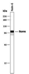
- Western blot analysis from lysates of Nalm-6 human Pre-B acute lymphocytic leukemia cell line. PVDF membrane was probed with 1 µg/mL of Goat Anti-human Ikaros Antigen Affinity-purified Polyclonal Antibody (Product # PA5-47682) followed by HRP-conjugated Anti-Goat IgG Secondary Antibody. Bands were detected for Ikaros (1-8 spice forms) at approximately 37 to 63 kDa (as indicated). This experiment was conducted under reducing conditions.

- Submitted by
- Invitrogen Antibodies (provider)
- Main image
- Experimental details
- Western blot analysis from lysates of Nalm-6 human Pre-B acute lymphocytic leukemia cell line. PVDF membrane was probed with 1 µg/mL of Goat Anti-human Ikaros Antigen Affinity-purified Polyclonal Antibody (Product # PA5-47682) followed by HRP-conjugated Anti-Goat IgG Secondary Antibody. Bands were detected for Ikaros (1-8 spice forms) at approximately 37 to 63 kDa (as indicated). This experiment was conducted under reducing conditions.

- Submitted by
- Invitrogen Antibodies (provider)
- Main image

- Experimental details
- Western blot analysis of IKAROS in Nalm‚6 human Pre‚B acute lymphocytic leukemia cell line. Samples were incubated in IKAROS polyclonal antibody (Product # PA5-47682) using a dilution of 1 µg/mL followed by a HRP-conjugated Anti-Goat IgG secondary antibody. Bands were detected for Ikaros (1-8 spice forms) at approximately 37 to 63 kDa (as indicated). This experiment was conducted under reducing conditions.

- Submitted by
- Invitrogen Antibodies (provider)
- Main image
- Experimental details
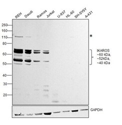
- Western blot was performed using Anti-IKAROS Polyclonal Antibody (Product # PA5-47682) and 60, 52 and 40 kDa band corresponding to IKZF1 along with an uncharacterised band (*) was observed across positive cell lines (REH, Daudi, Ramos and Jurkat) tested. Whole cell extracts (30 µg lysate) of Reh (Lane 1), Daudi (Lane 2), Ramos (Lane 3), Jurkat (Lane 4), U-937 (Lane 5), HL-60 (Lane 6), SH-SY5Y (Lane 7) and A-431 (Lane 8) were electrophoresed using NuPAGE™ 10% Bis-Tris Protein Gel (Product # NP0301BOX). Resolved proteins were then transferred onto a nitrocellulose membrane (Product # IB23001) by iBlot® 2 Dry Blotting System (Product # IB21001). The blot was probed with the primary antibody (1:200 dilution) and detected by chemiluminescence with Rabbit anti-Goat IgG (H+L) Superclonal™ Recombinant Secondary Antibody, HRP (Product # A27014,1:20000 dilution) using the iBright™ FL1500 Imaging System (Product # A44115). Chemiluminescent detection was performed using SuperSignal™ West Pico PLUS Chemiluminescent Substrate (Product # 34580).

- Submitted by
- Invitrogen Antibodies (provider)
- Main image

- Experimental details
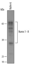
- Western blot analysis of IKAROS in 0.2 mg/mL lysates of Nalm‚6 human Pre-B acute lymphocytic leukemia cell line. Samples were incubated in IKAROS polyclonal antibody (Product # PA5-47682) using a dilution of 10 µg/mL followed by HRP-conjugated Anti-Goat IgG at a dilution of 0.0763888888888889. Specific bands were detected for Ikaros at approximately 63-77 kDa (as indicated). This experiment was conducted under reducing conditions and using the 12-230 kDa separation system.

- Submitted by
- Invitrogen Antibodies (provider)
- Main image
- Experimental details
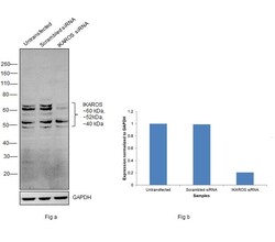
- Knockdown of IKZF1 was achieved by transfecting Jurkat with IKZF1 specific siRNAs (Silencer® select Product # s20179, s20180). Western blot analysis (Fig. a) was performed using Whole cell extracts from the IKZF1 knockdown cells (lane 3), non-targeting scrambled siRNA transfected cells (lane 2) and untransfected cells (lane 1). The blot was probed with IKAROS Polyclonal Antibody (Product # PA5-47682, 1:200 dilution) and Rabbit anti-Goat IgG (H+L) Superclonal™ Recombinant Secondary Antibody, HRP (Product # A27014,1:20000 dilution). Densitometric analysis of this western blot is shown in histogram (Fig. b). Decrease in signal upon siRNA mediated knock down confirms that antibody is specific to IKZF1.

Supportive validation
- Submitted by
- Invitrogen Antibodies (provider)
- Main image

- Experimental details
- Immunofluorescence analysis of IKZF1 was performed using log phase REH and HL60 cells. The cells were fixed with 4% paraformaldehyde for 10 minutes, permeabilized with 0.1% Triton™ X-100 for 15 minutes, and blocked with 2% BSA for 45 minutes at room temperature. The cells were labeled with IKAROS Polyclonal Antibody (Product # PA5-47682) at 1:100 in 0.1% BSA, incubated at 4 degree celsius overnight and then labeled with Rabbit anti-Goat IgG (H+L) Cross-Adsorbed Secondary Antibody, Alexa Fluor 488 (Product # A11078), (1:2000 dilution), for 45 minutes at room temperature (Panel a: Green). Nuclei (Panel b:Blue) were stained with ProLong™ Diamond Antifade Mountant with DAPI (Product # P36962). F-actin (Panel c: Red) was stained with Rhodamine Phalloidin (Product # R415, 1:300 dilution). Panel d represents the merged image showing nucleus and cytoplasm localization. Panel e represents HL60 which has no IKAROS expression. Panel f represents control cells with no primary antibody to assess background. The images were captured at 60X magnification.

Supportive validation
- Submitted by
- Invitrogen Antibodies (provider)
- Main image

- Experimental details
- Flow cytometric analysis of Jurkat human acute T cell leukemia cell line treated with 50 ng/mL PMA and 200 ng/mL calcium ionomycin for 30 minutes was fixed using formaldehyde, resuspended in lysis buffer, and sonicated to shear chromatin. Ikaros/DNA complexes were immunoprecipitated using 5 µg Goat Anti-human Ikaros Antigen Affinity-purified Polyclonal Antibody (Product # PA5-47682) or control antibody for 15 minutes in an ultrasonic bath, followed by Biotinylated Anti-Goat IgG Secondary Antibody. Immunocomplexes were captured using 50 µL of MagCellect Streptavidin Ferrofluid and DNA was purified using chelating resin solution. The VPAC promoter was detected by standard PCR.

- Submitted by
- Invitrogen Antibodies (provider)
- Main image

- Experimental details
- Flow cytometry of IKAROS in Jurkat human acute T cell leukemia cell line. Samples were incubated in IKAROS polyclonal antibody (Product # PA5-47682) or control antibody followed by Phycoerythrin-conjugated Anti-Goat IgG Secondary Antibody. To facilitate intracellular staining, cells were fixed with paraformaldehyde and permeabilized with methanol.

Supportive validation
- Submitted by
- Invitrogen Antibodies (provider)
- Main image

- Experimental details
- ChIP assay of IKAROS in Jurkat human acute T cell leukemia cell line. Samples were immunoprecipitated with IKAROS polyclonal antibody (Product # PA5-47682) for 15 minutes in an ultrasonic bath using a dilution of 5 μg followed by a Biotinylated Anti-Goat IgG secondary antibody. Cell line was treated with 50 ng/mL PMA and 200 ng/mL calcium ionomycin for 30 minutes was fixed using formaldehyde, resuspended in lysis buffer, and sonicated to shear chromatin. Immunocomplexes were captured using 50 μL of MagCellect Streptavidin Ferrofluid and DNA was purified using chelating resin solution. The VPAC promoter was detected by standard PCR.
