Explore
 Validate
 Learn
Learn Western blot
 Other assay
Antibody data
- Antibody Data
- Antigen structure
- References [1]
- Comments [0]
- Validations
- Other assay [2]
Submit
Validation data
Reference
Comment
Report error
- Product number
- PA5-27965 - Provider product page

- Provider
- Invitrogen Antibodies
- Product name
- DVL2 Polyclonal Antibody
- Antibody type
- Polyclonal
- Antigen
- Recombinant protein fragment
- Description
- Recommended positive controls: 293T, A431, H1299, HeLaS3, HepG2, Molt-4, Raji. Predicted reactivity: Mouse (99%), Rat (99%), Zebrafish (80%), Xenopus laevis (83%), Rhesus Monkey (99%), Bovine (96%). Store product as a concentrated solution. Centrifuge briefly prior to opening the vial.
- Reactivity
- Human, Mouse
- Host
- Rabbit
- Isotype
- IgG
- Vial size
- 100 µL
- Concentration
- 1.04 mg/mL
- Storage
- Store at 4°C short term. For long term storage, store at -20°C, avoiding freeze/thaw cycles.
Submitted references Islr regulates canonical Wnt signaling-mediated skeletal muscle regeneration by stabilizing Dishevelled-2 and preventing autophagy.
Zhang K, Zhang Y, Gu L, Lan M, Liu C, Wang M, Su Y, Ge M, Wang T, Yu Y, Liu C, Li L, Li Q, Zhao Y, Yu Z, Wang F, Li N, Meng Q
Nature communications 2018 Dec 3;9(1):5129
Nature communications 2018 Dec 3;9(1):5129
No comments: Submit comment
Supportive validation
- Submitted by
- Invitrogen Antibodies (provider)
- Main image

- Experimental details
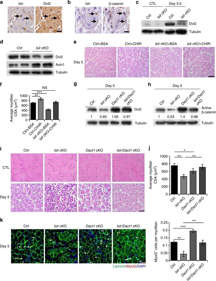
- Fig. 8 The ablation of Dact1 rescues the impaired skeletal muscle regeneration of Islr cKO mice by upregulating Dvl2. a Immunohistochemistry analysis of Islr and Dvl2 in injured TA muscles of WT mice at 5 d postinjury using serial sections. Scale bar = 10 mum. b Immunohistochemistry analysis of Islr and beta-catenin in injured TA muscles of WT mice at 5 d postinjury using serial sections. Scale bar = 10 mum. c Western blot analysis of Dvl2 protein levels in injured and CTL of control and Islr cKO mice at 3.5 d post injury. d Western blot analysis of Dvl2 and Axin1 protein levels in isolated satellite cells of control and Islr cKO mice cultured for 2 d in differentiation medium. e Intramuscular injection of CHIR or BSA at 2.5 d postinjury and H&E staining of injured TA muscles of control and Islr cKO mice at 5 d postinjury. N = 3 in each group. Scale bar = 100 mum . f CSA of myofibers in ( e ). g Western blot analysis of Dvl2 protein levels in injured TA muscles of control, Islr cKO, Dact1 cKO, and Islr / Dact1 cKO mice at 5 d postinjury. h Western blot analysis of active beta-catenin protein levels in injured TA muscles of control, Islr cKO, Dact1 cKO, and Islr / Dact1 cKO mice at 5 d postinjury. i H&E staining of injured and contralateral TA muscles in control, Islr cKO, Dact1 cKO, and Islr / Dact1 cKO mice at 5 d postinjury. Control (Ctrl): N = 5; Islr cKO: N = 3; Dact1 cKO: N = 3; Islr / Dact1 cKO: N =
- Submitted by
- Invitrogen Antibodies (provider)
- Main image

- Experimental details
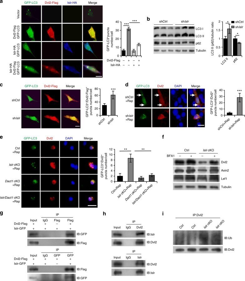
- Fig. 9 Islr stabilizes Dvl2 via antagonizing LC3-mediated autophagy. a Immunofluorescence analysis of GFP-LC3 + puncta in C2C12 cells transfected with GFP-LC3 and Dvl2-Flag plasmids, GFP-LC3 and Islr-HA plasmids, or GFP-LC3, Dvl2-Flag, and Islr-HA plasmids. N = 3 cell cultures in each group. Approximately, 20 cells total in each group. The numbers of GFP-LC3 + puncta per cell are shown on the right. b Western blot analysis of p62 and LC3II protein levels in shCtrl and sh Islr C2C12 cells after 2 d in growth medium. c Immunofluorescence analysis of GFP-LC3 + /Dvl2-Flag + puncta in shCtrl and sh Islr C2C12 cells transfected with GFP-LC3 and Dvl2-Flag plasmids. N = 3 cell cultures in each group. Approximately, 20 cells total in each group. The numbers of GFP-LC3 + /Dvl2-Flag + puncta per cell are shown on the right. d Immunofluorescence analysis of GFP-LC3 + /Dvl2 + puncta in shCtrl and sh Islr C2C12 cells treated with rapamycin (Rap) for 6 h. N = 3 cell cultures in each group. Approximately, 15 cells total in each group. The numbers of GFP-LC3 + /Dvl2 + puncta per cell are shown on the right. e Immunofluorescence analysis of GFP-LC3 + /Dvl2 + puncta in satellite cells from control, Islr cKO, Dact1 cKO, and Islr / Dact1 cKO mice treated with rapamycin (Rap) for 6 h. N = 3 cell cultures in each group. Approximately, 15 cells total in each group. The numbers of GFP-LC3 + /Dvl2 + puncta per cell are shown on the right. Scale bars are all 2