 Explore
 Validate
 Learn
Learn Western blot
Antibody data
- Antibody Data
- Antigen structure
- References [0]
- Comments [0]
- Validations
- Western blot [4]
- Immunocytochemistry [1]
Submit
Validation data
Reference
Comment
Report error
- Product number
- PA5-29244 - Provider product page

- Provider
- Invitrogen Antibodies
- Product name
- DVL2 Polyclonal Antibody
- Antibody type
- Polyclonal
- Antigen
- Recombinant protein fragment
- Description
- Recommended positive controls: 293T, A431, HeLa, HepG2, Jurkat, Raji, NCI-H929, Neuro2A, C8D30, NIH-3T3, Raw264.7, C2C12, PC-12, Rat-2. Predicted reactivity: Mouse (94%), Rat (92%), Rhesus Monkey (96%), Bovine (92%). Store product as a concentrated solution. Centrifuge briefly prior to opening the vial.
- Reactivity
- Human, Mouse, Rat
- Host
- Rabbit
- Isotype
- IgG
- Vial size
- 100 µL
- Concentration
- 1 mg/mL
- Storage
- Store at 4°C short term. For long term storage, store at -20°C, avoiding freeze/thaw cycles.
No comments: Submit comment
Supportive validation
- Submitted by
- Invitrogen Antibodies (provider)
- Main image

- Experimental details
- Western blot analysis of Dvl-2 using 30 µg of HCT116 lysate. Samples were loaded onto a 7.5% SDS-PAGE gel and probed with a Dvl-2 polyclonal antibody (Product # PA5-29244) at a dilution of 1:5000.

- Submitted by
- Invitrogen Antibodies (provider)
- Main image

- Experimental details
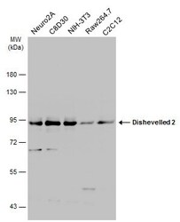
- Western Blot analysis of DVL2 was performed by separating 30 µg of various whole cell extracts by 7.5% SDS-PAGE. Proteins were transferred to a membrane and probed with a DVL2 Polyclonal Antibody (Product # PA5-29244) at a dilution of 1:5000 and a HRP-conjugated anti-rabbit IgG secondary antibody.

- Submitted by
- Invitrogen Antibodies (provider)
- Main image
- Experimental details
- Western Blot analysis of DVL2 was performed by separating 30 µg of various whole cell extracts by 7.5% SDS-PAGE. Proteins were transferred to a membrane and probed with a DVL2 Polyclonal Antibody (Product # PA5-29244) at a dilution of 1:5000 and a HRP-conjugated anti-rabbit IgG secondary antibody.

- Submitted by
- Invitrogen Antibodies (provider)
- Main image

- Experimental details
- Western Blot analysis of DVL2 was performed by separating 30 µg of various whole cell extracts by 7.5% SDS-PAGE. Proteins were transferred to a membrane and probed with a DVL2 Polyclonal Antibody (Product # PA5-29244) at a dilution of 1:5000 and a HRP-conjugated anti-rabbit IgG secondary antibody.

Supportive validation
- Submitted by
- Invitrogen Antibodies (provider)
- Main image
- Experimental details
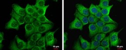
- Dvl-2 antibody [N1N3] detects Dvl-2 protein at cytoplasm by immunofluorescent analysis. Sample: HCT 116 cells were fixed in 4% paraformaldehyde at RT for 15 min. Green: Dvl-2 protein stained by Dvl-2 antibody [N1N3] (Product # PA5-29244) diluted at 1:500. Blue: Hoechst 33342 staining.
