Explore
 Validate
 Learn
Learn Western blot
 Immunocytochemistry
Antibody data
- Antibody Data
- Antigen structure
- References [5]
- Comments [0]
- Validations
- Immunocytochemistry [3]
- Immunohistochemistry [1]
- Flow cytometry [1]
- Other assay [4]
Submit
Validation data
Reference
Comment
Report error
- Product number
- PA5-71680 - Provider product page

- Provider
- Invitrogen Antibodies
- Product name
- SFTPC Polyclonal Antibody
- Antibody type
- Polyclonal
- Antigen
- Synthetic peptide
- Reactivity
- Human, Mouse, Rat
- Host
- Rabbit
- Isotype
- IgG
- Vial size
- 200 μL
- Concentration
- 0.5 mg/mL
- Storage
- Store at 4°C short term. For long term storage, store at -20°C, avoiding freeze/thaw cycles.
Submitted references Prkg2 regulates alveolar type 2-mediated re-alveolarization.
Interstitial lung disease in children with Rubinstein-Taybi syndrome.
Co-culture of type I and type II pneumocytes as a model of alveolar epithelium.
YAP and Wnt3a independently promote AECIIs proliferation and differentiation by increasing nuclear β‑catenin expression in experimental bronchopulmonary dysplasia.
Engineered amphiphilic peptides enable delivery of proteins and CRISPR-associated nucleases to airway epithelia.
Zhang M, Ali G, Komatsu S, Zhao R, Ji HL
Stem cell research & therapy 2022 Mar 21;13(1):111
Stem cell research & therapy 2022 Mar 21;13(1):111
Interstitial lung disease in children with Rubinstein-Taybi syndrome.
Bradford L, Ross MK, Minso J, Cernelc-Kohan M, Shayan K, Wong SS, Li X, Rivier L, Jegga AG, Deutsch GH, Vece TJ, Loughlin CE, Gower WA, Hurley C, Furman W, Stokes D, Hagood JS
Pediatric pulmonology 2022 Jan;57(1):264-272
Pediatric pulmonology 2022 Jan;57(1):264-272
Co-culture of type I and type II pneumocytes as a model of alveolar epithelium.
Brookes O, Boland S, Lai Kuen R, Miremont D, Movassat J, Baeza-Squiban A
PloS one 2021;16(9):e0248798
PloS one 2021;16(9):e0248798
YAP and Wnt3a independently promote AECIIs proliferation and differentiation by increasing nuclear β‑catenin expression in experimental bronchopulmonary dysplasia.
Jia X, Wu B, Huang J, Fan L, Yang M, Xu W
International journal of molecular medicine 2021 Jan;47(1):195-206
International journal of molecular medicine 2021 Jan;47(1):195-206
Engineered amphiphilic peptides enable delivery of proteins and CRISPR-associated nucleases to airway epithelia.
Krishnamurthy S, Wohlford-Lenane C, Kandimalla S, Sartre G, Meyerholz DK, Théberge V, Hallée S, Duperré AM, Del'Guidice T, Lepetit-Stoffaes JP, Barbeau X, Guay D, McCray PB Jr
Nature communications 2019 Oct 28;10(1):4906
Nature communications 2019 Oct 28;10(1):4906
No comments: Submit comment
Supportive validation
- Submitted by
- Invitrogen Antibodies (provider)
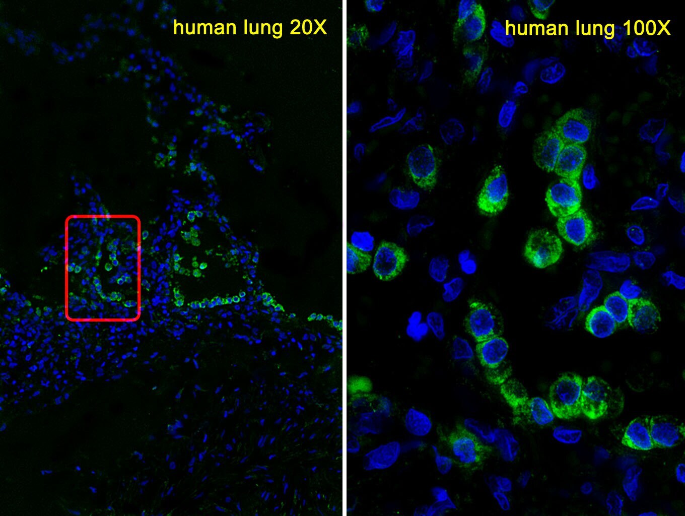
- Main image
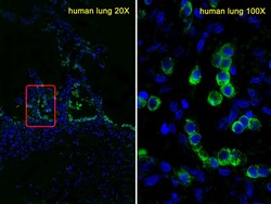
- Experimental details
- Immunofluorescent analysis of SFTPC in human lung cells using confocal microscopy. Samples were incubated with SFTPC H1 polyclonal antibody (Product # PA5-71680) using a dilution of 1:25. Alexa Fluor 488-conjugated goat anti-rabbit lgG (green) with a dilution of 1:400 was used as a secondary antibody. Nuclei were counterstained with DAPI (blue).
- Submitted by
- Invitrogen Antibodies (provider)
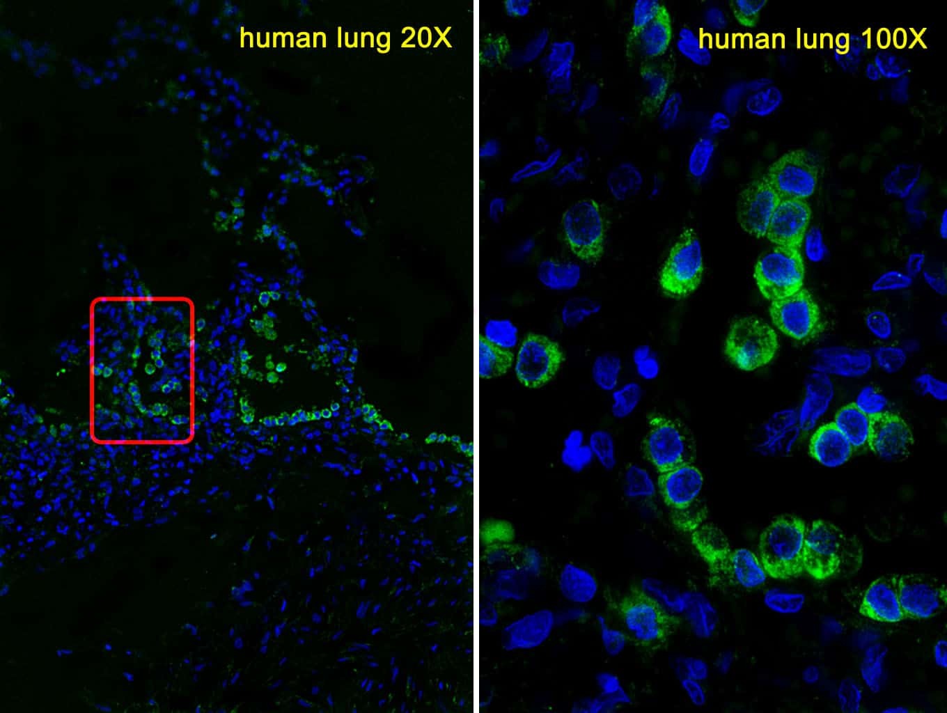
- Main image
- Experimental details
- Immunofluorescent analysis of SFTPC in human lung cells using confocal microscopy. Samples were incubated with SFTPC H1 polyclonal antibody (Product # PA5-71680) using a dilution of 1:25. Alexa Fluor 488-conjugated goat anti-rabbit lgG (green) with a dilution of 1:400 was used as a secondary antibody. Nuclei were counterstained with DAPI (blue).
- Submitted by
- Invitrogen Antibodies (provider)
- Main image
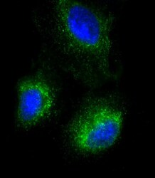
- Experimental details
- Immunocytochemistry analysis of SFTPC in A549 cells. Samples were incubated with SFTPC polyclonal antibody (Product # PA5-71680) using a dilution of 1:25 followed by Dylight® 488-conjugated goat anti-Rabbit IgG at a dilution of 1:200 (green). Cells were 4% paraformaldehyde-fixed, 0. 1% Triton X-100 permeabilized. Immunofluorescence image showing cytoplasm staining on A549 cell line. Cytoplasmic actin is detected with Dylight® 554 Phalloidin at 1:500 dilution (red). The nuclear counter stain is DAPI (blue).
Supportive validation
- Submitted by
- Invitrogen Antibodies (provider)
- Main image
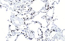
- Experimental details
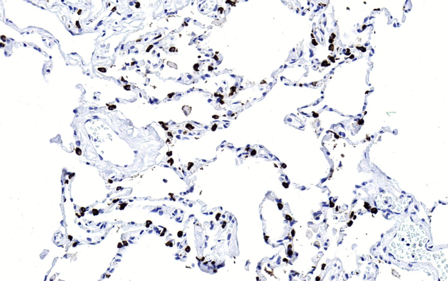
- Immunohistochemistry analysis of SFTPC in paraffin-embedded Human lung section. Samples were incubated with SFTPC polyclonal antibody (Product # PA5-71680) using a dilution of 1:200 followed by an undiluted biotinylated goat polyvalent antibody was used as the secondary, followed by DAB staining.
Supportive validation
- Submitted by
- Invitrogen Antibodies (provider)
- Main image

- Experimental details
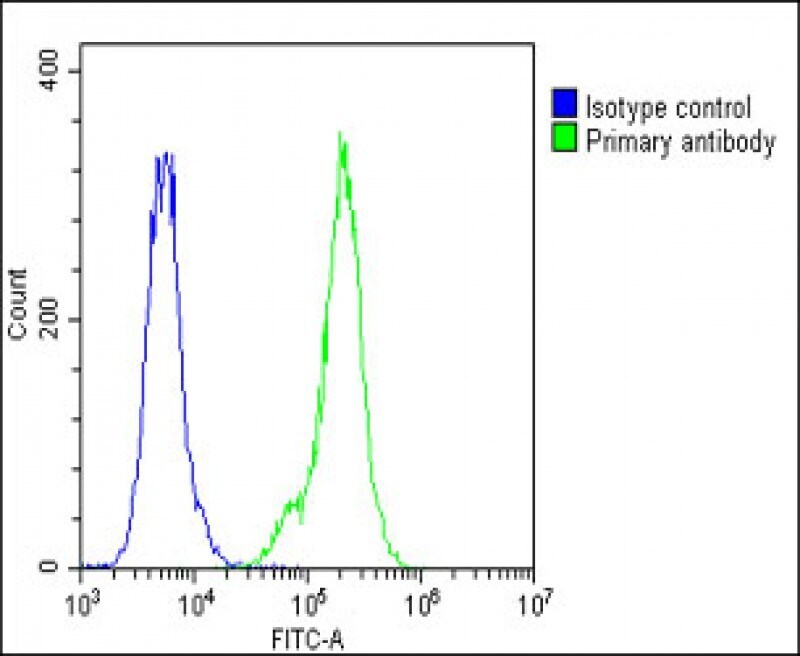
- Flow cytometry of (overlay histogram) of SFTPC in A549 cells (green line). Samples were incubated with SFTPC polyclonal antibody (Product # PA5-71680) using a dilution of 1:25 dilution for 60 min at 37°C followed by Goat-Anti-Rabbit IgG, DyLight® 488 Conjugated Highly Cross-Adsorbed at 1:200 dilution for 40 min at 37°C. The cells were fixed with 2% paraformaldehyde (10 min) and then permeabilized with 90% methanol for 10 min. The cells were then incubated in 2% bovine serum albumin to block non-specific protein-protein interactions followed by the primary antibody. Isotype control antibody (blue line) was rabbit IgG1 (1 μg/1x10^6 cells) used under the same conditions. Acquisition of >10, 000 events was performed.
Supportive validation
- Submitted by
- Invitrogen Antibodies (provider)
- Main image
- Experimental details
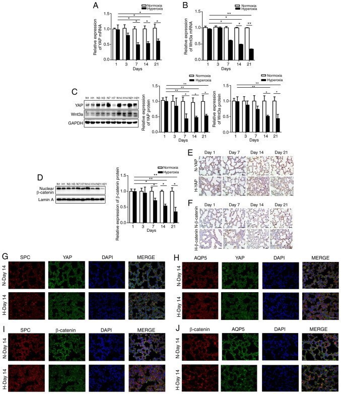
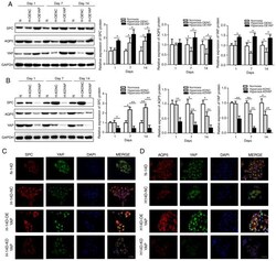
- Figure 2 YAP, Wnt3a and beta-catenin expression is decreased in lung tissue affected by BPD. (A) YAP mRNA and (C) protein expression in the lungs. (B) Wnt3a mRNA and (C) protein expression in the lungs. (D) beta-catenin protein expression in lung nucleoli. Data are expressed as the means +- SD (n=6). * P
- Submitted by
- Invitrogen Antibodies (provider)
- Main image
- Experimental details
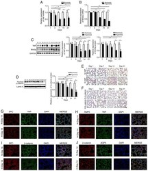
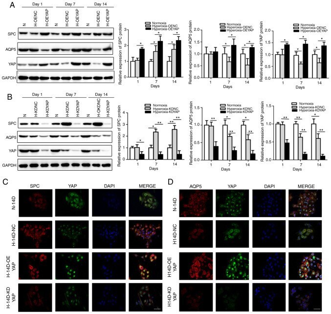
- Figure 3 Effects of YAP on the proliferation and differentiation of AECIIs into AECIs. (A and B) SPC and AQP5 expression in OEYAP, KDYAP and corre-sponding control (OENC, KDNC) AECIIs detected by western blot analysis. Data are expressed as the means +- SD (n=3). * P
- Submitted by
- Invitrogen Antibodies (provider)
- Main image
- Experimental details
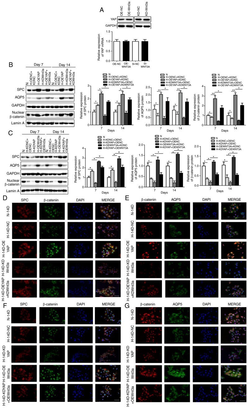
- Figure 5 Wnt3a overexpression and knockdown reversed the effects of YAP on AECIIs proliferation and differentiation. (A) YAP expression in OEWnt3a, KDWnt3a and corresponding control (OENC, KDNC) AECIIs determined by western blot analysis. Data are expressed as the means +- SD (n=3). (B, D and E) SPC, AQP5 and nuclear beta-catenin expression detected by (B) western blot analysis and double IF staining for (D) SPC and beta-catenin or (E) AQP5 and beta-catenin in OEYAP, KDWnt3a, OEYAP + KDWnt3a, and corresponding control (OENC, KDNC) AECIIs extracted from hyperoxia-exposed rats after 14 days. Data are expressed as the means +- SD (n=3). * P
- Submitted by
- Invitrogen Antibodies (provider)
- Main image
- Experimental details
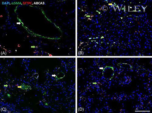
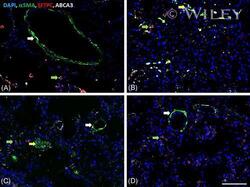
- 3 Figure Immunofluorescence overlay images; x20; blue (DAPI): nuclei; Green (FITC): alpha-smooth muscle actin (aSMA); Red (AF568): surfactant protein C (SP-C); white (AF633): ATP binding cassete-A3 (ABCA3) (A) control; (B) Case 1; (C) Case 2; (D) Case 3. Green arrows: SP-C and ABCA3 colocalization in alveolar type 2 (AT2) pneumocytes; white arrows: aSMA staining around small vessels and airways; yellow arrows: aSMA+ interstitial myofibroblasts [Color figure can be viewed at wileyonlinelibrary.com ]