 Explore
 Validate
 Learn
Learn Western blot
Antibody data
- Antibody Data
- Antigen structure
- References [1]
- Comments [0]
- Validations
- Western blot [2]
- Immunocytochemistry [1]
- Other assay [1]
Submit
Validation data
Reference
Comment
Report error
- Product number
- 711292 - Provider product page

- Provider
- Invitrogen Antibodies
- Product name
- FKBP5 Recombinant Polyclonal Antibody (6HCLC)
- Antibody type
- Polyclonal
- Antigen
- Synthetic peptide
- Reactivity
- Human, Mouse, Rat
- Host
- Rabbit
- Isotype
- IgG
- Antibody clone number
- 6HCLC
- Vial size
- 100 µg
- Concentration
- 0.5 mg/mL
- Storage
- Store at 4°C short term. For long term storage, store at -20°C, avoiding freeze/thaw cycles.
Submitted references Endothelial Cell-Specific Transcriptome Reveals Signature of Chronic Stress Related to Worse Outcome After Mild Transient Brain Ischemia in Mice.
Wegner S, Uhlemann R, Boujon V, Ersoy B, Endres M, Kronenberg G, Gertz K
Molecular neurobiology 2020 Mar;57(3):1446-1458
Molecular neurobiology 2020 Mar;57(3):1446-1458
No comments: Submit comment
Supportive validation
- Submitted by
- Invitrogen Antibodies (provider)
- Main image

- Experimental details
- Western blot analysis was performed on whole cell extracts (30 µg lysate) of HeLa (Lane 1), HT-29 (Lane 2), HCT 116 (Lane 3), HEL (Lane 4), HEK-293 (Lane 5), Jurkat (Lane 6) and K-562 (Lane 7). The blots were probed with Anti-FKBP5 Recombinant Rabbit Polyclonal Antibody (Product # 711292, 1-2 µg/mL) and detected by chemiluminescence using Goat anti-Rabbit IgG (H+L) Superclonal™ Secondary Antibody, HRP conjugate (Product # A27036, 0.4 µg/mL, 1:2500 dilution). A 51 kDa band corresponding to FKBP5 was observed across cell lines tested. Known quantity of protein samples were electrophoresed using Novex® NuPAGE® 4-12% Bis-Tris gel (Product # NP0321BOX), XCell SureLock™ Electrophoresis System (Product # EI0002) and Novex® Sharp Pre-Stained Protein Standard (Product # LC5800). Resolved proteins were then transferred onto a nitrocellulose membrane with iBlot® Dry Blotting System (Product # IB21001). The membrane was probed with the relevant primary and secondary Antibody following blocking with 5% skimmed milk. Chemiluminescent detection was performed using Pierce™ ECL Western blotting Substrate (Product # 32106).

- Submitted by
- Invitrogen Antibodies (provider)
- Main image

- Experimental details
- Knockdown of FKBP5 was achieved by transfecting HeLa cells with FKBP5 specific siRNAs (Silencer® select Product # s5215). Western blot analysis (Fig. a) was performed using whole cell extracts from the FKBP5 knockdown cells (lane 3), non-specific scrambled siRNA transfected cells (lane 2) and untransfected cells (lane 1). The blot was probed with FKBP5 Recombinant Rabbit Polyclonal Antibody (Product # 711292, 1:1000 dilution) and Goat anti-Rabbit IgG (H+L) Superclonal™ Secondary Antibody, HRP conjugate (Product # A27036, 0.25µg/ml, 1:4000 dilution). Densitometric analysis of this Western blot is shown in histogram (Fig. b). Decrease in signal upon siRNA mediated knock down confirms that antibody is specific to FKBP5.

Supportive validation
- Submitted by
- Invitrogen Antibodies (provider)
- Main image

- Experimental details
- Immunofluorescence was performed on fixed and permeabilized HeLa cells for detection of endogenous FKBP5 using Anti-FKBP5 Recombinant Rabbit Polyclonal Antibody (Product # 711292, 2 µg/mL) and labeled with Goat anti-Rabbit IgG (H+L) Superclonal™ Secondary Antibody, Alexa Fluor® 488 conjugate (Product # A27034, 1:2000). Panel a) shows representative cells that were stained for detection and localization of FKBP5 protein (green), Panel b) is stained for nuclei (blue) using SlowFade® Gold Antifade Mountant with DAPI (Product # S36938). Panel c) represents cytoskeletal F-actin staining using Alexa Fluor® 555 Rhodamine Phalloidin (Product # R415, 1:300). Panel d) is a composite image of Panels a, b and c clearly demonstrating nuclear localization of FKBP5. Panel e) represents control cells with no primary antibody to assess background.

Supportive validation
- Submitted by
- Invitrogen Antibodies (provider)
- Main image
- Experimental details
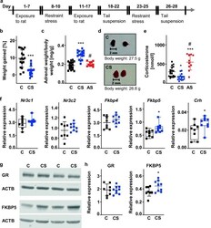
- Fig. 1 Characterization of the neuroendocrine effects of the chronic stress paradigm. a Schematic diagram of experimental design. b Body weight was recorded before the beginning and at the end of the chronic stress procedure. Chronically stressed mice gain less body weight than unstressed control animals. C: n = 18, CS: n = 17. Unpaired t test. t = 5.262, *** p < 0.001. c , d Chronic stress increases the weight of the adrenal glands. C: n = 18, CS: n = 17, AS: n = 10. One-way ANOVA F(2,42) = 40.14, p < 0.001 with Tukey's multiple comparison test: *** p < 0.001 CS versus C, # p < 0.001 CS versus AS. e Corticosterone plasma levels were measured at the beginning of the light cycle. C: n = 18, CS: n = 17, AS: n = 10. One-way ANOVA F(2,42) = 26.20, p < 0.001 with Tukey's multiple comparison test: * p < 0.05 CS versus C, *** p < 0.001 AS versus C, # p < 0.001 CS versus AS. f Hypothalamic mRNA transcription of genes associated with corticosteroid signaling. Nr3c1 , nuclear receptor subfamily 3 group C member 1. Nr3c2 , nuclear receptor subfamily 3 group C member 2. Fkbp4 , FK506 binding protein 4. Fkbp5 , FK506 binding protein 5. Crh , corticotropin releasing hormone. Relative mRNA expression is reported as the value normalized to tripeptidyl peptidase 2 ( Tpp2 ) for each sample. C: n = 8, CS: n = 9. Mann-Whitney U test. Nr3c1 : U = 27, p = 0.423. Fkbp5: U = 12, *p < 0.05. Crh: U = 14, * p < 0.05. Unpaired t test . Nr3c2 : t = 1.016, p = 0.326. Fkbp4 : t = 1.214, p = 0.244. g Repres
